< Back
Intensifier Control
Revealing Cancer's Infrastructure

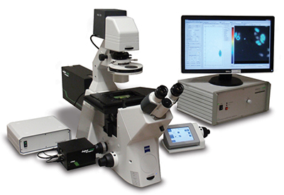
This year marks the 10th anniversary of the LIFA. With the first Lambert Instruments FLIM Attachment (LIFA) a decade ago, we introduced an easy and fast approach to fluorescence lifetime imaging. Since then, we advanced our imaging and analysis software; we improved our hardware and made it more compact; and we added compatibility with third-party hardware. But at the heart of the LIFA experience are still the features that matter most to our users. They are using the LIFA every day, because it is the easiest and fastest system for fluorescence lifetime imaging microscopy.
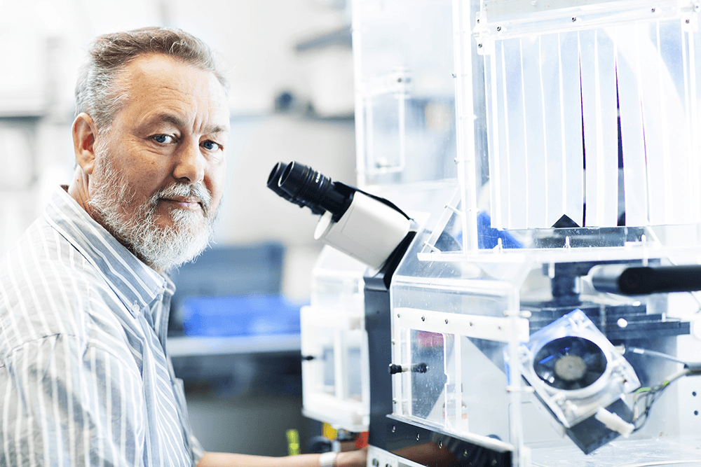
We visited Dr. Kees Jalink of the Biophysics of Cell Signaling group at the Netherlands Cancer Institute. His research group purchased the first ever LIFA to leave the labs of Lambert Instruments. Ten years later, the LIFA is still their fluorescence lifetime imaging method of choice for studying signal transduction pathways in living cells.
“Cancer can only be truely understood by knowing it in great detail,” says Jalink, “that is, by knowing exactly where and when signal transduction pathways become activated. Tools to study signals must yield data with spatial and temporal detail from living cells, preferably from cells that are as much as possible in a natural environment.”
“We use FRET sensors to study living cells. So for us, some of the other fluorescence lifetime imaging methods are just unacceptably slow. Our researchers are always in a hurry, because the cells they want to study expire within a couple of hours. The LIFA allows us to quickly record fluorescence lifetime images and immediately see a visualization of the results. It is the only way to quickly gather quantitative FLIM data.”


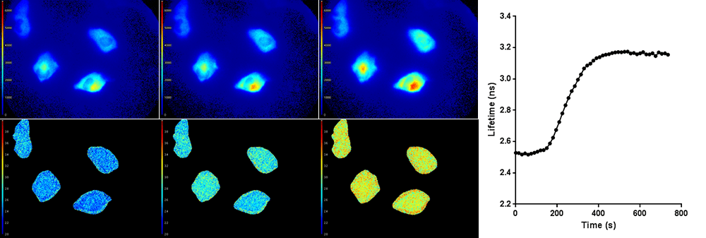
Top row: Light intensity images (colorized). Bottom row: Corresponding fluorescence lifetime images (colorized). The average fluorescence lifetime of the cells increases over time, as shown in the graph on the right.
All researchers and students in Jalink’s group are trained to work with the LIFA to analyze their cells. “After an introduction of about 30 minutes, everybody can work with the system. It’s a simple procedure and that’s why we use the LIFA nearly every day.”
More Information
Lambert Instruments has been shipping the LIFA to cancer research facilities all over the world for years. We are hopeful that we have facilitated a major advance in cancer research. If you are in this field of research, please contact us if you have any questions about the LIFA.
Publications
Over the years, many researchers have published the results of experiments they performed with the LIFA. We have compiled an overview of LIFA publications, as well as a number of application notes that illustrate some of its many applications.


