< Back
with SPAD powered vTAU camera
The complete high-speed recording solution
High-speed camera for fluorescence imaging
Intensified high-speed camera
Intensified camera with ultra-short gating
High-speed Intensified Camera Attachment
Compact lens-coupled image intensifier
Intensifier Control
Unit for Automated Systems
Record and edit high-speed videos with one or multiple cameras.
Record and analyze fluorescence lifetime images.
Custom imaging products, sensors and software for low light level applications.

Confocal FLIM Applications

Confocal imaging on a widefield fluorescence microscope can now be done in combination with frequency-domain fluorescence lifetime imaging microscopy (FLIM). The increased spatial resolution in the z-direction results in lifetime images with enhanced contrast as the detection of out-of-focus emission is reduced significantly. This allows you to see differences in fluorescence lifetime e.g. between the cell membrane and the cytoplasm.

The data below were obtained with the Lambert Instruments FLIM Attachment (LIFA), either widefield (with LED light; 468nm peak) or confocal (with spinning disk CSU10 and 470nm-diode laser). The fluorescence lifetime images are generated at 2 different z-positions, z1 and z2. Snapshots of several z-positions are shown, as well as movies through even more.

Z1

Z2

CONFOCAL
(SPINNING DISK)

WIDEFIELD
(LED)

These images show different pollen grains: the lifetime in pseudo colours and the intensity in grey scale. Because of the pinholes in the spinning disk, the exposure time is higher with confocal imaging. In this image 220 ms exposure time per phase step was taken for the confocal image versus 195 ms for the widefield (LED light). However, when a diode laser with higher power is used, the exposure time can be shortened.

Z1

Z2

CONFOCAL
(SPINNING DISK)

WIDEFIELD
(LED)

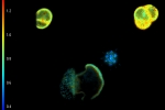
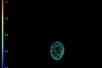
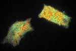
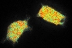
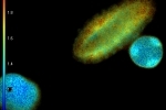
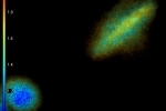
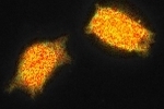
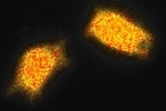
These images show YFP-transfected mammalian cells and were taken with 12 phase steps of each 400 ms for the confocal, versus 70 ms for the widefield image. The calculated lifetimes are 2.34 ns (z1) and 2.42 ns (z2) in the confocal images, versus 2.58 ns (z1) and 2.57 ns (z2) in the widefield images.

The differences in lifetime could be due to the fact that the diode laser has one excitation wavelength, exactly 470 nm, while the LED has a range of wavelengths for which we used the emission band pass filter of 465-495 nm.

Contact Us

5th floor,
Leonard Springerlaan 19
9727KB Groningen
The Netherlands

Phone:
+31 (0) 50 501 8461
Email:
sales@lambertinstruments.com