Custom imaging products, sensors and software for low light level applications.
TIRF FLIM
Total Internal Reflection Fluorescence (TIRF) microscopy is a super-resolution technique with high sensitivity of fluorescence near the cover glass. TIRF does not disturb cellular activity, and enables tracking of biomolecules, and the study of their dynamic activity and interactions at the molecular level. TIRF enables the selective visualisation of processes and structures of the cell membrane and pre-membrane space such as vesicle release and transport, cell adhesion, secretion, membrane protein dynamics and distribution or receptor-ligand interactions. The combination of TIRF and frequency domain FLIM makes it possible to measure fluorescence lifetimes of for instance small focal adhesions near the cover glass.
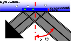
High NA TIRF objectives (up to 1.49) make it possible to introduce illumination at incident angles greater than the critical angle (θ ) resulting in TIR (Total Internal Reflection) accompanied by the formation of an evanescent wave immediately adjacent to the coverglass-specimen interface. The evanescent wave energy drops off exponentially with distance from the coverglass and reaches about a hundred nanometers into the specimen. For TIR to occur, the refractive index of the coverglass should be higher than the refractive index of the specimen (which is e.g. the case when using buffered saline solution). The white-TIRF as well as the laser-TIRF system utilises this evanescent wave to excite fluorescent molecules in a very thin section in contact with the coverglass (here: green dots). Because the specimen is not excited beyond the evanescent wave (here: white dots), this imaging system can produce fluorescence images with an extremely high signal-to-noise (S/N) ratio and z-resolution.