< Back
with SPAD powered vTAU camera
The complete high-speed recording solution
High-speed camera for fluorescence imaging
Intensified high-speed camera
Intensified camera with ultra-short gating
High-speed Intensified Camera Attachment
Compact lens-coupled image intensifier
Intensifier Control
Unit for Automated Systems
Record and edit high-speed videos with one or multiple cameras.
Record and analyze fluorescence lifetime images.
Custom imaging products, sensors and software for low light level applications.

Total Internal Reflection Fluorescence Lifetime Imaging Microscopy

Total Internal Reflection Fluorescence (TIRF) microscopy facilitates extremely high-contrast visualization and thereby high sensitivity of fluorescence near the cover glass. Typically, the optical section adjacent to the cover glass is about 100 nm. TIRF does not disturb cellular activity, thus enabling tracking of biomolecules, and the study of their dynamic activity and interactions at the molecular level. TIRF enables the selective visualisation of processes and structures of the cell membrane and pre-membrane space like vesicle release and transport, cell adhesion, secretion, membrane protein dynamics and distribution or receptor-ligand interactions. The unique combination of TIRF and frequency domain FLIM makes it possible to measure lifetimes of, for instance, small focal adhesions near the cover glass.

TIRF

WIDEFIELD

LIFETIME (PSEUDOCOLORS)

INTENSITY (GRAYSCALE)

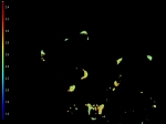
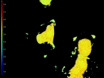
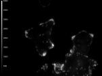
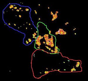
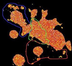
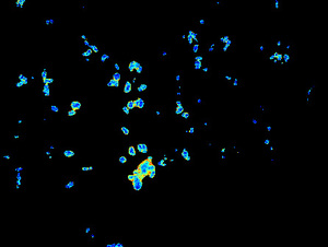
These cells (kindly provided by Ms. S.E. Le Devedec, Leiden University, The Netherlands) express dSH2-GFP in small focal adhesions as well as in the nuclei as shown by widefield microscopy. However, by the use of TIRF only fluorescence close to the coverslip is obtained, thus only the focal adhesions are excited.

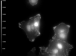
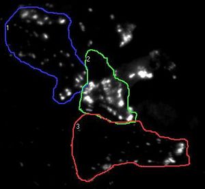
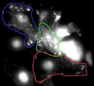
Fluorescence lifetime images give a more accurate measurement in TIRF mode, as out of focus light is emitted from the average lifetime in the focal adhesions.

The images shown here are taken with the Nikon TE2000-U widefield microscope with white-TIRF illuminator, combined with the Lambert Instruments Fluorescence lifetime imaging Attachment (LIFA). As light source the modulated LED of 468nm 3W was used and as demonstrated here enough intensity was generated to obtain fluorescence lifetime images with TIRF.

TIRF

WIDEFIELD

LIFETIME (PSEUDOCOLORS)

INTENSITY (GRAYSCALE)

Cells (kindly provided by Ms. S.E. Le Devedec, Leiden University, The Netherlands) expressing dSH2-GFP in small focal adhesions as well as in the nuclei.

The images shown here are taken with the Olympus TIRFM (laser-TIRF), combined with the Lambert Instruments FLIM Attachment (LIFA). As light source the modulated diode laser of 473 nm 20 mW was used and as demonstrated here enough intensity was generated to obtain fluorescence lifetime images with TIRF.

TIRF

LIFETIME (PSEUDOCOLORS)

INTENSITY (GRAYSCALE)

Cells (kindly provided by Ms. S.E. Le Devedec, Leiden University, The Netherlands) expressing dSH2-GFP in small focal adhesions as well as in the nuclei.

Contact Us

5th floor,
Leonard Springerlaan 19
9727KB Groningen
The Netherlands

Phone:
+31 (0) 50 501 8461
Email:
sales@lambertinstruments.com