

In the realm of modern scientific exploration and medical diagnostics, fluorescence imaging stands out as a powerful technique, offering unparalleled insights into the world of cells, molecules, and tissues. From unraveling the mysteries of biological processes to advancing medical diagnostics, it has revolutionized the way researchers and clinicians visualize and understand the intricate workings of life.
Understanding Fluorescence Imaging
It is a versatile imaging technique that utilizes the natural fluorescence properties of certain molecules or artificially introduced fluorophores to generate detailed, high-resolution images. This technique relies on the principle of fluorescence, wherein certain substances absorb light at a specific wavelength and then emit light at a longer wavelength.
Applications in Scientific Research
In scientific research, it plays a crucial role in elucidating various biological phenomena. Researchers use fluorescent markers or dyes to label specific molecules or structures within cells, allowing them to track molecular interactions, observe cellular processes, and study the localization of proteins with remarkable precision.
From studying the dynamics of gene expression to investigating the behavior of individual cells in complex biological systems, fluorescence imaging provides researchers with a powerful tool to explore the intricacies of life at the molecular level.
Advancements in Medical Diagnostics
In the field of medical diagnostics, it offers a non-invasive and highly sensitive approach for detecting and visualizing diseased tissues. Fluorescent contrast agents can be selectively targeted to specific biomarkers or pathological features, enabling clinicians to identify tumors, visualize blood vessels, and assess tissue viability with exceptional accuracy.
Cutting-Edge Technologies
Recent advancements in fluorescence imaging technologies have further expanded its capabilities and applications. High-speed cameras, advanced microscopy techniques, and sophisticated image analysis algorithms have enhanced the resolution, sensitivity, and speed of fluorescence imaging, enabling researchers and clinicians to delve deeper into the intricacies of biological system

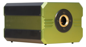
Cooled High-speed Camera for Fluorescence Imaging
Intensified high-speed high-sensitivity imaging camera
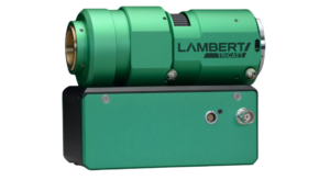
Compact lens-coupled image intensifier
Future Directions
As this type of imaging continues to evolve, researchers are exploring new avenues and pushing the boundaries of what is possible. Emerging technologies such as super-resolution microscopy, multiphoton imaging, and optogenetics promise to further revolutionize our understanding of cellular dynamics and disease mechanisms.
Conclusion
Fluorescence imaging represents a cornerstone of modern scientific research and medical diagnostics, offering unprecedented insights into the inner workings of life. With its versatility, sensitivity, and non-invasive nature, fluorescence imaging continues to drive discovery and innovation across a wide range of disciplines. As we look to the future, the potential of fluorescence imaging to uncover new mysteries and transform healthcare remains boundless.
For more information click here: https://en.wikipedia.org/wiki/Fluorescence_imaging