< Back
Intensifier Control
Ion imaging
For ion imaging, several fluorescent indicators (sensor, construct, tracer, etc) are available that have a change in quantum yield upon ion binding. This means that they emit photons with different energy, thus have different emission wavelength. Their fluorescence lifetime could also change. Therefore, there are two methods in which ion imaging can be done by use of indicators: the ratiometric method and the FLIM method.
Another method ion imaging is by the use of Forster Resonance Energy Transfer based (FRET-based) indicators that change their conformation upon ion binding. Upon the conformational change of a FRET-based indicator, its FRET efficiency changes, which is used as indicator of ion concentration. Examples of these indicators are cameleons. Cameleons are genetically-encoded fluorescent indicators for Ca2+ based on green fluorescent protein variants and calmodulin (CaM).
Reference: Miyawaki A, Griesbeck O, Heim R, Tsien RY. “Dynamic and quantitative Ca2+ measurements using improved cameleons”. Proc Natl Acad Sci USA (PNAS) 96(5):2135-40 (1999)
CALCIUM IMAGING
Calcium (Ca2+) is important for signal transduction pathways.
PROTON (PH) IMAGING
The intracellular proton (H+) concentration (pH), as well as intracellular calcium, is important in the regulation of cellular functions including growth, differentiation, motility, exocytosis and endocytosis. To study this in more detail, measurements of the intracellular pH of resting cells can be done and the pH fluctuations inside cells after environmental perturbations can be followed.
Reference: Hai-Jui Lin, Petr Herman, and Joseph R. Lakowicz. “Fluorescence Lifetime-Resolved pH Imaging of Living Cells”. Cytometry Part A 52A:77–89 (2003).

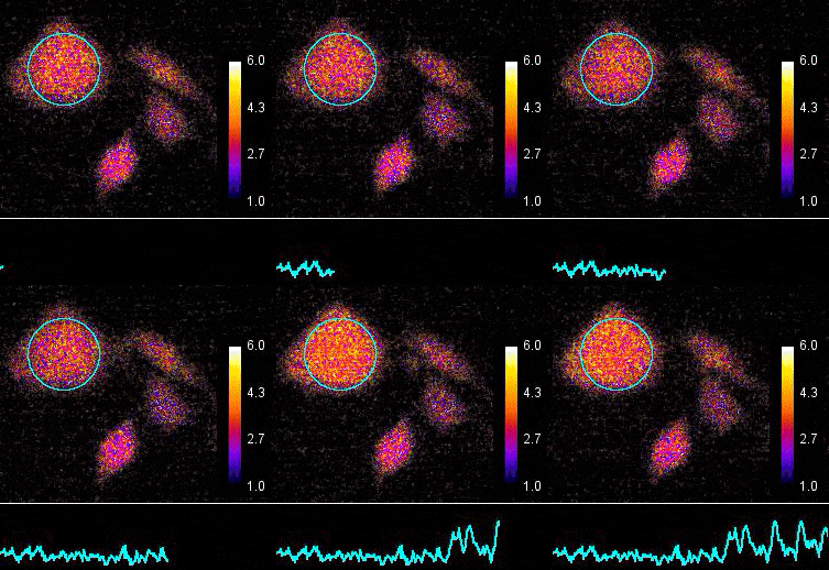
Demonstration of the Lambert Instruments Toggel camera for single-image FLIM (siFLIM) detection of histamine-induced alterations in Ca2+ concentration. Tiny oscillations in Ca2+ levels (~2.5 s periods) are observed after addition of histamine. Such small and rapid transients would go completely unnoticed when recorded by conventional FLIM.
Video courtesy of the Netherlands Cancer Institute.
CALCIUM IMAGING
Calcium (Ca2+) is important for signal transduction pathways.
PROTON (PH) IMAGING
The intracellular proton (H+) concentration (pH), as well as intracellular calcium, is important in the regulation of cellular functions including growth, differentiation, motility, exocytosis and endocytosis. To study this in more detail, measurements of the intracellular pH of resting cells can be done and the pH fluctuations inside cells after environmental perturbations can be followed.
Reference: Hai-Jui Lin, Petr Herman, and Joseph R. Lakowicz. “Fluorescence Lifetime-Resolved pH Imaging of Living Cells”. Cytometry Part A 52A:77–89 (2003).
ZINC IMAGING
Zinc (Zn2+) is involved in enzyme catalysis, protein structure, protein-protein interactions, and protein-oligonucleotide interactions. Zinc interacts with extracellular binding sites, which are likely to include binding sites involved in the subsequent translocation of this ion to the cell interior. Inside the cell, Zinc binds to cytosolic and organelle binding sites or is taken up by intracellular organelles.
SODIUM IMAGING
Sodium (Na+) is important in the signal transduction in the central nerve system.
MAGNESIUM IMAGING
Many enzymes (like kinases) require the presence of magnesium Mg2+ ions for their catalytic action, especially enzymes utilising ATP.
CHLORIDE IMAGING
Chloride (Cl-) plays a role in the central nervous system.
POTASSIUM
Potassium (K+) plays a role in cell growth and cell viability.
INDICATORS FOR ION IMAGING BY FLIM
BCECF (pH)
Bis-BTC (heavy metals)
Calcium-crimson (Calcium, orange excitation)
Calcium-green (Calcium, blue excitation)
Carboxyfluorescein (pH)
Carboxy-SNAFL-1 (pH)
Carboxy-SNAFL-2 (cytosol pH)
DM-NERF dextrans (lysosoml pH)
Fluo-3 (Calcium)
Fura-2 (Calcium)
LysoSensor DND-160 (lysosomal pH)
LysoSensor probe (pH)
Magnesium-green (Magnesium)
Mag-quin-1 (Magnesium)
Mag-quin-2 (Magnesium)
MQAE (Chloride)
Newport Green DCF (Zinc)
OG-514 carboxylic acid dextrans (lysosoml pH)
PBFI (Potassium)
Quin-2 (Calcium, blue excitation)
SPQ (Chloride)


