

Cell biology is the discipline that studies cells to answer scientific questions. All organisms are composed of one or more cells and all vital functions of an organism occur within cells. Cells contain the hereditary information necessary for regulating cell functions. Cells possess DNA, the hereditary material of genes, and RNA, containing the information necessary to build various proteins such as enzymes, the cell’s primary machinery. There are also other kinds of biomolecules in cells, e.g. lipids, proteins, macromolecules, and more.
Cell biology research includes learning the physiological properties such as the structure and the organelles of cells, their environment, interactions, life cycle, division, function, and eventual death. This is done both on microscopic and molecular level, and includes the research of single-celled organisms like bacteria as well as specialized cells in multi-cellular organisms like humans.
Knowing the composition of cells and how cells work is fundamental to all of the biological sciences. Appreciating the similarities and differences between cell types is particularly important to the fields of cell and molecular biology. These fundamental similarities and differences provide a unifying theme, allowing the principles learned from studying one cell type to be extrapolated and generalized to other cell types.
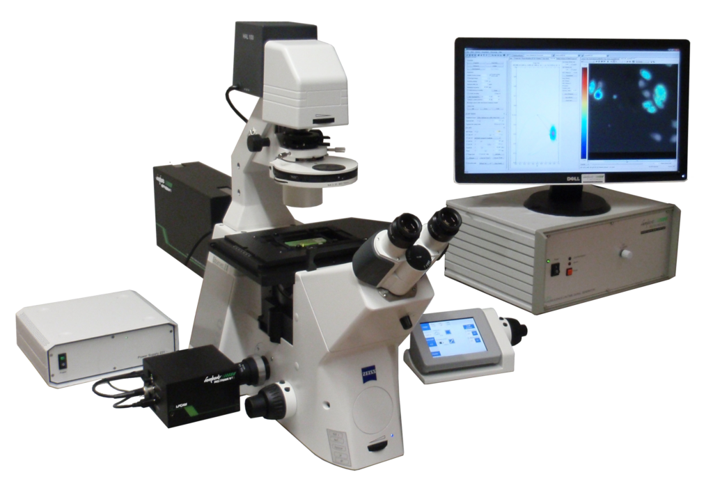
In combination with our products, wide field fluorescence microscopy is used to measure characteristics of fluorescent proteins. Cells, originating from bacteria and insects to mammals, generally are kept in culture and plated at coverslips to do specific experiments. Microscopy enables viewing objects inside cells that are stained or fluorescently tagged. By observing the characteristics, e.g. the fluorescence lifetime, of the fluorescent compounds, not just the localization of a fluorescent protein, but also the characteristics of its local environment can be imaged. Novel multi-parameter fluorescence imaging systems are being used to study intracellular organization and inter- and intracellular signalling.
One way to observe the proteins, is by fixation of the cells to the coverslips. Before the cells are fixed, the compounds in the cells can be fluorescently tagged (see living cell-imaging). Also, the compounds inside the cells can be stained after their fixation, for example by use of antibodies. Staining is a biochemical technique of adding a class-specific (DNA, proteins, lipids or carbohydrates) dye to a substrate to qualify or quantify the presence of a specific compound. The characteristics of the dyes can give answers to specific scientific questions, like whether there is interaction between two different proteins, whether there is a conformational change of the protein after a kind of treatment (see also Fluorescence Lifetime Imaging Microscopy and Forster Resonance Energy Transfer), or whether specific ions have bound to the proteins of interest, etc.
The cells can also be analyzed in-vivo. These living cell imaging experiments seek to gain information about the localization and interaction of the desired protein. One way to do this is to replace the wild-type gene with a ‘fusion’ gene that has a reporting element such as GFP. That will allow easy visualization of the products of the genetic modification. More sophisticated techniques are in development that can track protein products without mitigating their function, such as the addition of small sequences which will serve as binding motifs to monoclonal antibodies.

5th floor,
Leonard Springerlaan 19
9727KB Groningen
The Netherlands