< Back
with SPAD powered vTAU camera
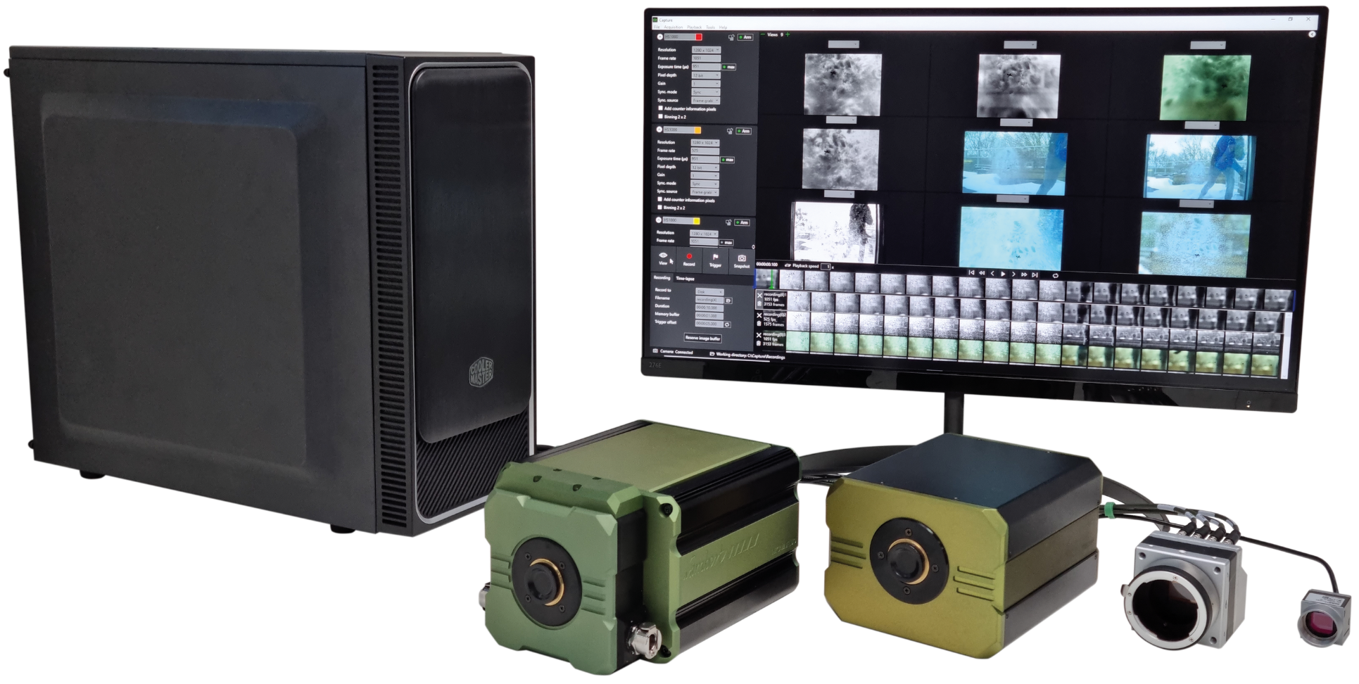
The complete high-speed recording solution
High-speed camera for fluorescence imaging
Intensified high-speed camera
Intensified camera with ultra-short gating
High-speed Intensified Camera Attachment
Compact lens-coupled image intensifier
Intensifier Control
Unit for Automated Systems
Record and edit high-speed videos with one or multiple cameras.
Record and analyze fluorescence lifetime images.
Custom imaging products, sensors and software for low light level applications.

LIFA FLIM System

with SPAD powered vTAU camera

LIFA FLIM

Product Overview

The Lambert Instruments LIFA FLIM system is the fastest and easiest way to perform Fluorescence Lifetime Imaging Microscopy (FLIM).

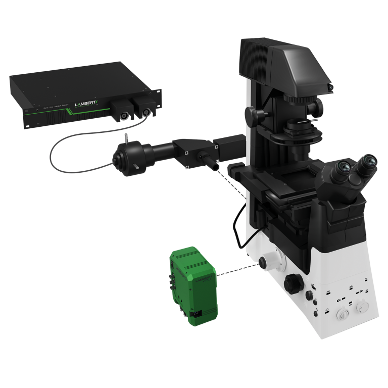
Available in versatile configurations dependent on your specific applications, the LIFA system offers a turn-key solution for fluorescence lifetime imaging microscopy. Compatible with any fluorescence microscope with a camera output – including microscopes by Leica, Nikon, Olympus, TILL and Zeiss. Set up is quick and easy, with all hardware integrated seamlessly with our dedicated LIFA software, so you can focus on your experiment.

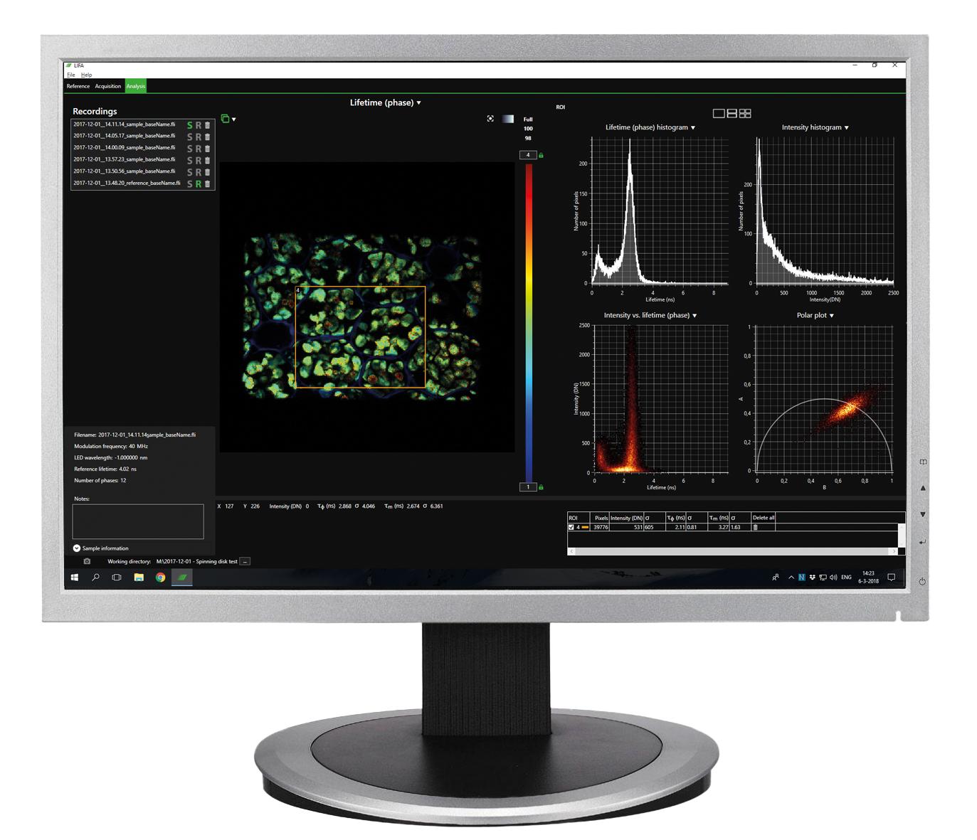
The advanced software instantly analyses data and presents the calculated fluorescence lifetimes visually. Recorded images are compatible with ImageJ, FIJI, Matlab and MetaMorph, while detailed statisitcal data can be exported to an Excel worksheet.

Read more about LIFA software | Watch our FLIM webinar.

Captured with LIFA vTAU: Dual-color, high-speed 3D imaging of blood flow in a beating embryonic zebrafish heart.

Applications

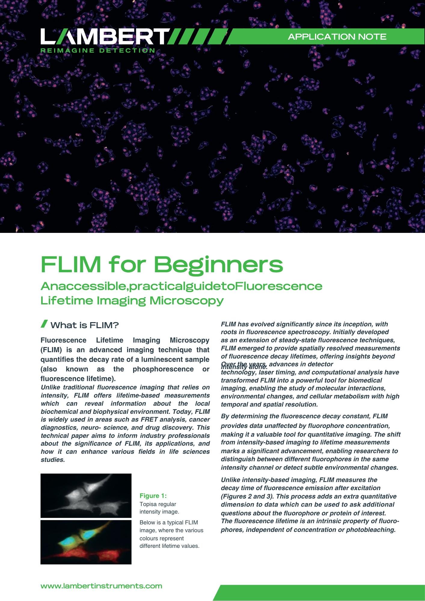
FLIM for Beginners Application Note

Features

Experiments from start to finish

From recording images to lifetime calculation and data analysis

Dedicated LIFA software

Using our stand-alone LIFA Software, you can instantly calculate the fluorescence lifetime and presents it as a colour coded overlay and a phaser plot.

Broad Lifetime Range

From sub microsecond down to picoseconds

Comprehensive SDK

Our software development kit (SDK) provides a flexible platform for integrating and automating experiments, offering you full control and customization of your fluorescence lifetime measurements.

Multiple configurations

System components to suit your applications

Non-phototoxic illumination

Configurations

Widefield

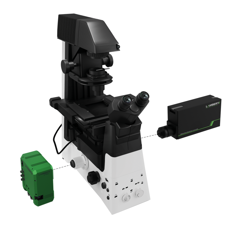
The Lambert Instruments VTAU camera, when combined with the Multi-LED, provides a compact and effective solution for FLIM on widefield microscopes.

The VTAU easily connects to the camera port, while the Multi-LED integrates with the standard epifluorescence port, making the setup compatible with most widefield microscope configurations.

Spinning-disk confocal

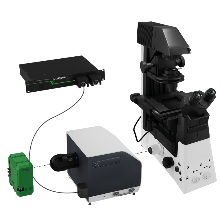
The Lambert Instruments LIFA VTAU system for frequency-domain FLIM is well-suited for camera-based multi-beam confocal techniques.

It is compatible with systems such as the Yokogawa CSU series (Nipkow disk), Andor Dragonfly, Crest V3/V2/Cicero, and Visitech International’s VTInfinity series, enabling versatile imaging performance.

TIRF

Total Internal Reflection Fluorescence (TIRF) microscopy enables high-contrast fluorescence imaging close to the cover glass, typically within a ~100 nm optical section.

When combined with frequency-domain FLIM, it allows for lifetime imaging of structures such as membrane receptors, aiding in the analysis of cellular signalisation pathways.

Featured LIFA users

System Components

The LIFA VTAU system includes:

LIFA Software

Seamless integration of hardware for full system control

Light Sources

Multi-LED and/or Multi-LASER as required for your set up

OEM Solution

We offer an OEM platform based on the proven Lambert Instruments LIFA system

Frequently Asked Questions

Fluorescence Lifetime Imaging Microscopy (FLIM) is a technique that measures the decay time of fluorescence molecules, so-called fluorophores. FLIM provides detailed information about molecular environments, energy transfer between fluorophores and protein interactions. It is widely used in biological and biomedical research to study dynamic processes and cellular microenvironments.

Time-domain Fluorescence Lifetime Imaging Microscopy (FLIM) is a technique that measures the fluorescence lifetime of molecules by exciting fluorophores with a very short pulse of light and then detecting the time it takes for them to emit photons. The emitted light’s intensity decays over time, and this decay curve is analyzed to determine the fluorescence lifetime.

Frequency-domain Fluorescence Lifetime Imaging Microscopy (FLIM) is a technique used to measure the fluorescence lifetimes of fluorophores by modulating the excitation light at different frequencies. In this method, the phase shift and modulation depth of the emitted light relative to the excitation source are measured, which provides information about the fluorescence lifetime. This technique is valuable for studying molecular interactions, environment-sensitive probes, and cellular dynamics. FLIM is commonly used in biological and biomedical imaging, to sense the cellular environment, get a better understanding of cell biology or to differentiate between fluorophores based on their lifetimes if they have similar emission spectra.

The primary difference between frequency-domain and time-domain FLIM lies in how the fluorescence lifetime is measured:

Frequency-domain FLIM: Modulates the excitation light at specific frequencies and measures the phase shift and modulation of the emitted light relative to the excitation. It determines lifetime from these shifts.

Time-domain FLIM: Uses short light pulses to excite fluorophores and measures the time delay between the excitation pulse and the emitted photons. The fluorescence decay curve is analyzed to determine lifetime.

Both techniques provide insights into molecular environments but differ in how they capture fluorescence data.


Widefield microscopy is an imaging technique used in biological and clinical research to capture images of entire specimens or large sections of tissue. It involves illuminating the entire field of view at once with light, typically from a broad light source, such as a lamp or LED. All parts of the sample are illuminated simultaneously, allowing for the detection of light emitted or reflected from the entire specimen. This method is commonly used for fluorescence microscopy, enabling researchers to observe labeled structures in live or fixed cells.

Spinning-disk confocal microscopy is an advanced imaging technique used for capturing high-resolution images of biological samples in real-time. It employs a rotating disk with multiple pinholes to focus light onto specific areas of the sample while rejecting out-of-focus light. This enables faster image acquisition compared to traditional laser-scanning confocal microscopy. Spinning-disk confocal is ideal for live-cell imaging due to its speed, lower phototoxicity, and reduced photobleaching, making it suitable for dynamic processes like cell movement or protein interactions.

Total Internal Reflection Fluorescence (TIRF) microscopy is a technique used to observe events occurring near the surface of a cell, typically at or near the plasma membrane. TIRF systems use an evanescent wave generated by light undergoing total internal reflection at the interface between two media (e.g., glass and water) to selectively illuminate and excite fluorophores within a very thin region, typically less than 200 nm, adjacent to the surface. This minimizes background noise and allows high-contrast imaging of cellular processes like protein interactions at the cell membrane.

Light sheet microscopy, also known as Selective Plane Illumination Microscopy (SPIM), is a technique used to image large, three-dimensional biological samples with minimal phototoxicity and photobleaching. In this method, a thin sheet of light illuminates a single plane of the sample, while an orthogonal detection system captures high-resolution images. By moving the light sheet through the sample, researchers can reconstruct detailed 3D images of tissues, embryos, or entire organisms. This method is especially useful for long-term live imaging of biological processes in real-time.

Request More Information

    Lambert Instruments BV

    Leonard Springerlaan 19
    9727 KB Groningen
    The Netherlands

    TRiCAM

    Intensified low-light imaging CMOS camera with ultra-short gating

    COMPACT AND COST EFFECTIVE

    10-20 times more light efficiency in a compact and light weight form

    HIGHLY CUSTOMISABLE

    choose from a wide variety of high sensitivity image intensifiers to match the needs of your application

    SINGLE-PHOTON SENSITIVE

    high sensitivity supplemented with an acquisition speed up to 160 fps.

    Description

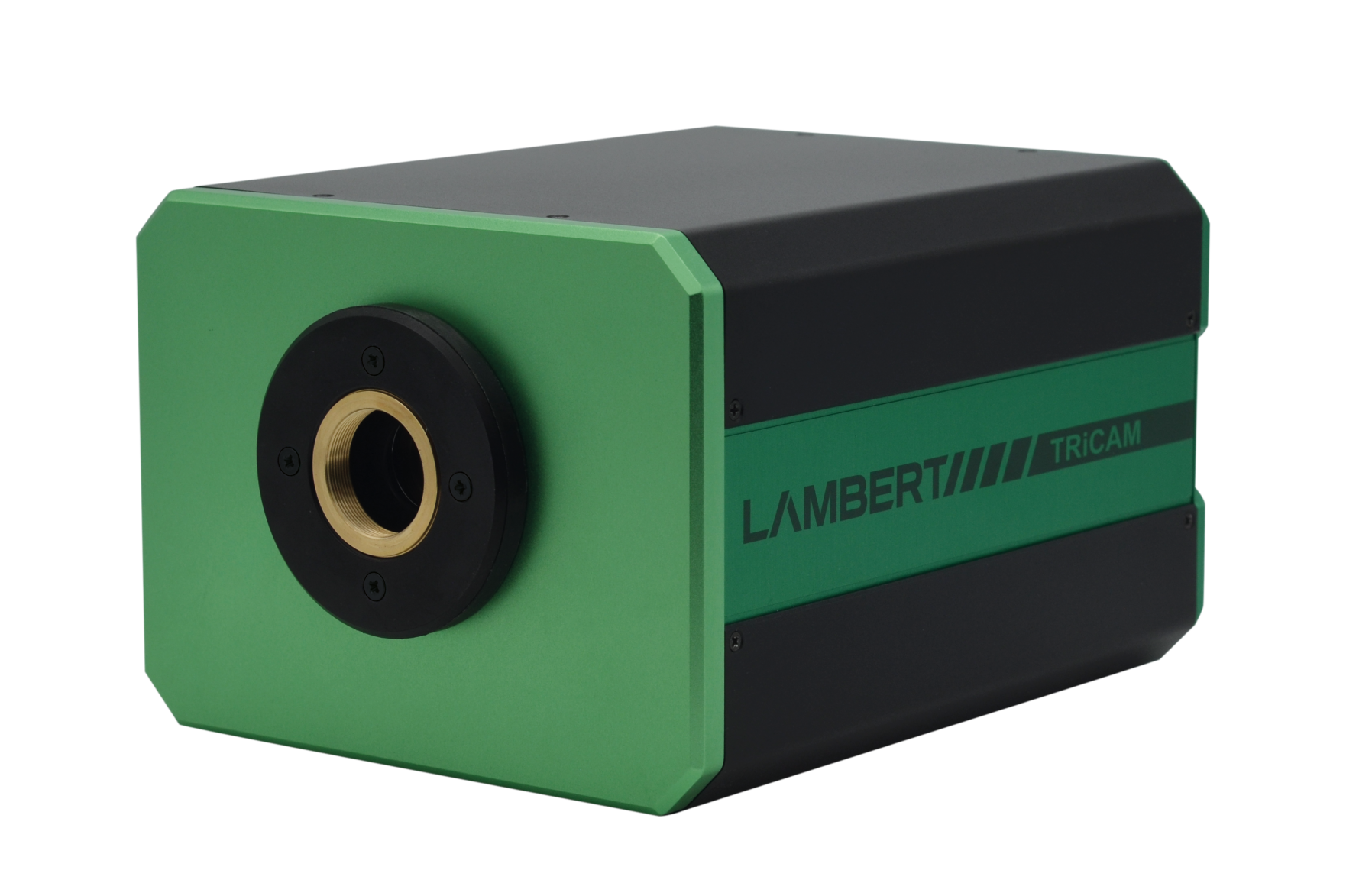
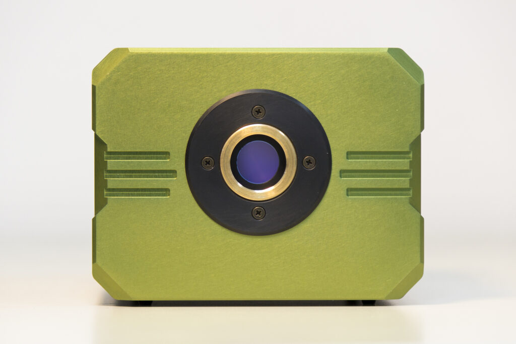
    The TRiCAM is a compact intensified low-light imaging CMOS camera. It is designed for scientific and industrial applications that require low-light imaging. With built-in signal generators, the TRiCAM is capable of ultra-short exposures through fast gating and therefore suitable for time-resolved imaging.

    Applications

    Laser Induced Fluorescence (LIF)

    Time-gated luminescence

    Bio- and chemiluminescence imaging

    Plasma physics

    Single Photon imaging

    Particle Image Velocimetry (PIV)

    Solar PV and LED characterization

    Combustion research

    Single-molecule imaging

    Fluorescence Lifetime-Imaging

    Microscopy (FLIM)

    Förster Resonance Energy Transfer

    (FRET)

    Time-gated Raman / Laser Induced

    Breakdown Spectroscopy (LIBS)

    Time-resolved imaging & spectroscopy

    Diffuse Optical Tomography (DOT)

    High voltage Partial Breakdown (Partial discharge)

    TRiCAM CMOS Camera

    Features

    Gated image intensifier
    The TRiCAM is equipped with an integrated timing pulse generator and a gate-unit, generating gate pulses down to < 3 ns.

    Low-light imaging
    The TRiCAM has a built-in image intensifier that boosts the incoming light. This way, you can capture detailed images in the most challenging light conditions.

    Fiber-optically coupled
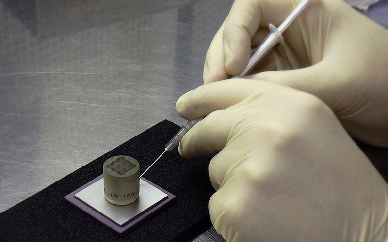
    Our experienced engineers couple the sensor to the image intensifier with a fiber-optic plate, resulting in a 10 – 20x more light efficient system. The fiber-optic plate is a solid piece of glass that consists of millions of parallel glass fibers sealed together. Each fiber acts as an independent light conductor that transfers the light from the image intensifier to the sensor.

    Ultra-short gating
    The image intensifier in the TRiCAM can be used as an ultra-fast electronic shutter. This technique is called gating and it can be done in a matter of nanoseconds.

    Gating can eliminate motion blur when imaging fast-moving objects or highly dynamic processes. By varying the timing of the gate signal very precisely (tens of picosecond resolution), you can use gating to record a time-resolved light intensity profile.

    Optimised for your application
    The TRiCAM can be configured with a wide range of image intensifiers. Available image intensifiers cover the entire visual spectrum and the near infrared.

    High-resolution sensor
    The TRiCAM features a high-resolution CMOS sensor. It captures stunningly detailed images at 1920 x 1200 pixels.

    160 fps
    At up to 160 fps, the TRiCAM can record slow-motion footage, even in low-light conditions.

    Global shutter
    With its global shutter readout method, the sensor in the TRiCAM eliminates rolling shutter effects in your images.

    Fluorescence Imaging

    Image Intensifiers 

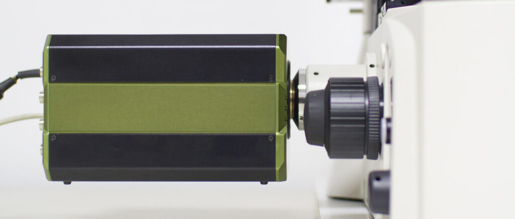
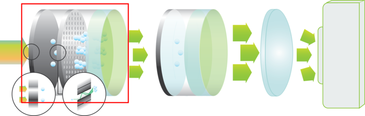
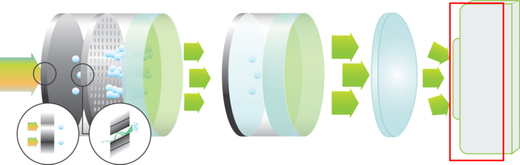
    An image intensifier boosts the intensity of the incoming light. By converting photons to electrons and back to photons, the light intensity can be increased significantly. Scroll down for more information about how this works.

    Another feature of an image intensifier is that it can act as an ultra-fast shutter. The photocathode can be switched between on and off in a matter of nanoseconds.

    Read More

    Low-light imging 

    The TRiCAM has a built-in image intensifier that boosts the incoming light. This way, you can capture detailed images in the most challenging light conditions.

    Our experienced engineers will help you pick the right image intensifier for your application.

    Fluorescence Imaging

    Photocathodes 

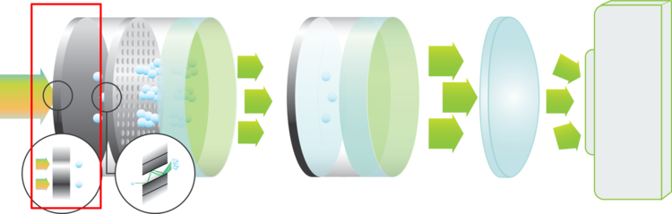
    The photocathode is the entrance of the image intensifier. This is where the incoming photons are converted to electrons. The quantum efficiency of the photocathode material specifies how efficient this conversion is for each wavelength.

    Read More

    Images

    TRiCAM CMOS Camera
    TRiCAM CMOS Camera

    Specifications

    IMAGE INTENSIFIER

    Diameter: 18 mm

    Minimum Gate Width: 40 ns (< 3 ns optional)

    Maximum Repetition Frequency: 300 kHz

    Trigger Input: TTL

    SENSOR

    Resolution: 1920 x 1200 pixels

    Pixel Size: 5.86 µm

    Frame Rate: 162 fps

    Sensor Type: CMOS

    Readout Method: Global Shutter

    ADC: 10 bit and 12 bit

    Downloads

    For more information on the TRiCAM CMOS Camera, please download the brochure.

    Request more information

      Lambert Instruments BV

      Leonard Springerlaan 19
      9727 KB Groningen
      The Netherlands

      User Publications

      Researchers around the world use the TRiCAM in their studies. Opposite is an overview of the most recent publications describing research that was done using a TRiCAM.

      For a complete overview of applications, please visit our applications pages.

      Control Unit

      Intensifier Control for automated systems

      COMPACT

      and possibility to request for custom firmware

      EFFICIENCY

      Computer controlled by USB connection

      SIMPLICITY

      Seamlessly integrates with Lambert Instruments’ intensifier attachments

      Description

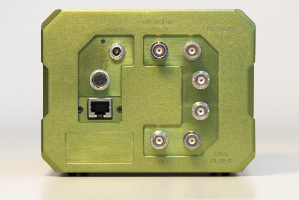
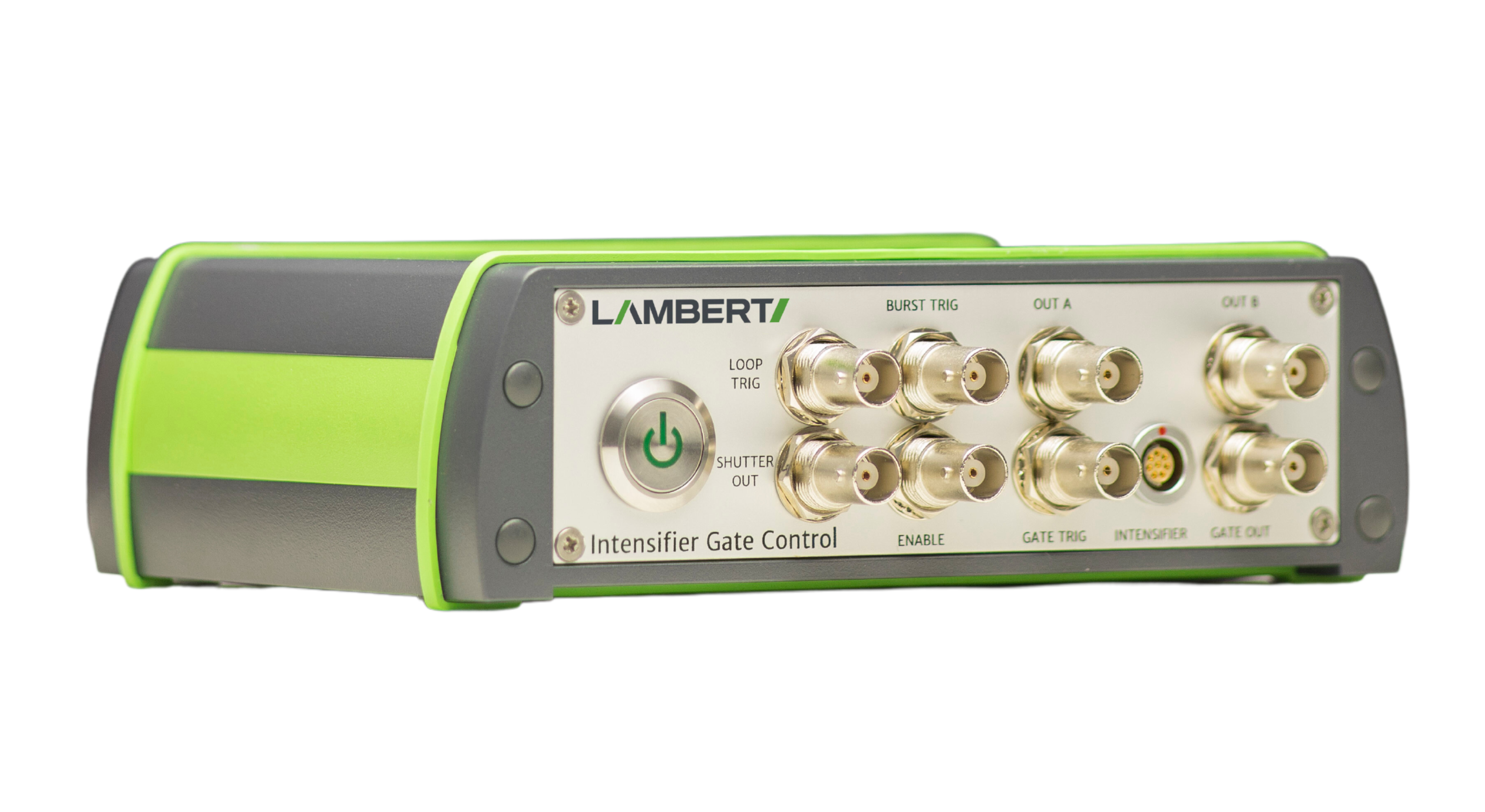
      For integration into a larger system or for automated measurements, the gain control unit for automated systems lets you control the settings of the intensifier attachment. The control unit uses standard TTL and analog signals for communication, allowing the user to switch the intensifier on or off, alter the gain and the anode current limit of the image intensifier without the need of software integration.


      Applications

      Time resolved imaging

      Synchronization imaging

      Rapid Gate switching to improve dynamic range

      To be used with Lambert Instruments’ Intensifier Attachments.

      Features

      Image Intensifier Gain
      By increasing the image intensifier gain, the incoming light intensity will be boosted more, resulting in a brighter image. The intensifier control for automated systems lets you control the gain of the image intensifier.

      Anode Current Limit
      To protect the fragile image intensifier from being damaged by overexposure, the anode current limiter can be used to set a limit for the acceptable anode current. If the anode current exceeds this value, then the image intensifier will be switched to a safe mode.

      Compatibility
      The gain control unit for automated systems is compatible with our intensifier attachments: HiCATT and TRiCATT

      Specifications

      A synchronisation signal is needed in order to create an output at the three outputs of the intensifier gate control. This signal can originate from different sources:

      External source: For instance a signal from a frame grabber or a connected camera.

      Software command

      Internal sync source: Gives synchronisation pulses with a fixed frequency. Can generate pulses at frequencies between 0.5 Hz and 4 MHz.

      When using an external synchronisation source, the following modes are available:

      – Synchronise with rising edge of external input pulse

      – Synchronise with falling edge of external input pulse

      – Follow external input pulse

       
      Advanced gating modes (can be customised to a further extent) 
       
      – Burst mode
      – Multiple exposures per sync pulse
      – Multiple exposures and Burst mode
      – Rapid gate-setting switching 
      – Enable gate
      – Precision gating
      Timing Resolution 250ps
      Jitter Pulse Width <250 ps RMS
      Jitter Delay <250 ps RMS
      Sync with falling/rising edge Follow external output post
      Pulse width /delay range 10ns – 10s 10ns – 10s
      Timing resolution (Step size) 10ns 10ns
      Jitter pulse width <10ps RMS <10ps RMS
      Jitter delay <10ns RMS <250ns RMS
      Insertion delay  30ns 20ns

      Request more information

        Lambert Instruments BV

        Leonard Springerlaan 19
        9727 KB Groningen
        The Netherlands

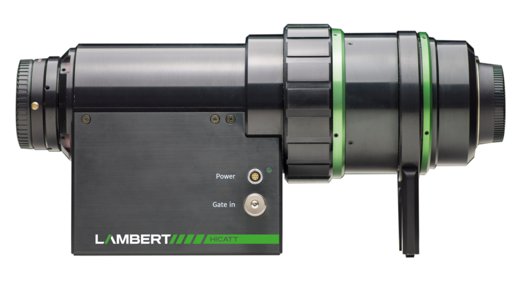
        HiCATT

        High-speed camera attachment for intensified imaging

        HiCATT Image Intensifier

        10 MHz IN
        BURST FOR MORE THAN 250 FRAMES

        for low light imaging with increased sensitivity 

        CYCLED
        BURSTS

        adjust for changing light levels by cycling through three different gate widths

        IN-SITU FOCUS ADJUSTMENT

        outer-body focus ring adjusts the internal relay optics without altering total length

        Description

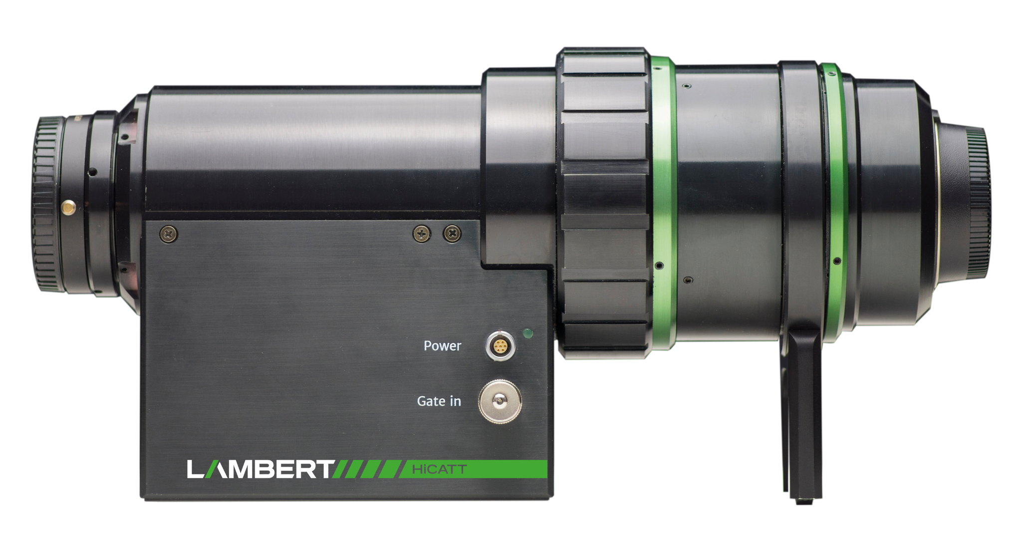
        The High-speed Intensified Camera Attachment (HiCATT) is designed for use with a high-speed camera. It increases the sensitivity of your camera and enables low-light imaging at frame rates up to 1 MHz (10 Mhz in burst)

        The technology in the HiCATT expands the dynamic range of your high-speed camera. At low light levels, even single photons can be detected. While at high light levels, overexposure can be prevented by very short exposures (down to 3 ns). These short exposures yield sharp images of fast moving objects.


        Applications

        Combustion research / Hydrogen discovery 

        High-speed fluorescence, bioluminescence and chemiluminescence detection for in-vivo imaging

        Super-slow motion combustion research for the automotive industry

        Time-resolved imaging in plasma physics research

        Dynamic phenomena in microscopy

        Laser-Induced Fluorescence (LIF)

        Many other industrial or scientific low-light-level applications in high-speed imaging

        UV imaging for hydrogen and fusion

        HiCATT High-Speed Camera Attachment

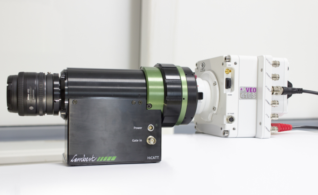
        The HiCATT is an intensifier attachment for your high-speed camera. It provides increased light sensitivity for high-speed imaging applications. In this photo, the HiCATT is attached to a Phantom Veo 410S.

        Features

        Optimised for your application
        The HiCATT can be configured with a wide range of image intensifiers. Our experienced engineers will help you pick the right image intensifier for your application.

        High-speed Imaging
        The HiCATT upgrades your high-speed camera to the next level of performance. It boosts the intensity of incoming light at speeds up to 1,000,000 fps.

        Ultra-short exposures
        The gated image intensifier enables exposure times down to 3 ns. At such short exposure times, motion blur is eliminated completely to ensure sharp images.

        50% QE high-sensitivity intensifiers
        You can choose from a wide variety of high-sensitivity image intensifiers to match the spectral needs of your application.

        Rotating camera mount
        Safe and easy camera coupling keeping the HiCATT and the camera in their intended orientation.

        Compatible with Shimadzu and Vision Research – We can provide fully integrated solutions tailored to your setup.

        Match my camera

        The relay optics of HiCATT and TRiCATT project the output of the image intensifier onto the sensor of your camera. This calculator helps you determine the optimal configuration of image intensifer diameter and relay optics for your camera and application.

        Read More

        Fluorescence Imaging

        Image Intensifiers 

        An image intensifier boosts the intensity of the incoming light. By converting photons to electrons and back to photons, the light intensity can be increased significantly. Scroll down for more information about how this works.

        Another feature of an image intensifier is that it can act as an ultra-fast shutter. The photocathode can be switched between on and off in a matter of nanoseconds.

        Read More

        Fluorescence Imaging

        Photocathodes 

        The photocathode is the entrance of the image intensifier. This is where the incoming photons are converted to electrons. The quantum efficiency of the photocathode material specifies how efficient this conversion is for each wavelength.

        Read More

        Phosphors

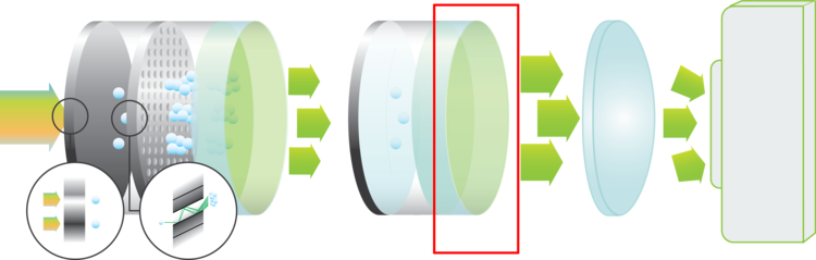
        The output of the image intensifier contains a layer of phosphorescent material. Upon impact of an electron, the phosphor screen will emit light. Depending on the type of phosphor, the intensity of the emitted light will decrease faster.

        Videos

        Butane-Propane Flame at 4200 FPS
        Flames (mix Butane – Propane) at 4200 fps and 40 us gate open time (effective exposure time), HiCATT 25 image intensifier, high-speed camera attachment with Phantom V4.0 high-speed camera.

        Electronic Discharge at 47000 FPS
        Electronic Discharge at 47000 fps and 3 us gate open time (effective exposure time), HiCATT 25 image intensifier, high-speed camera attachment with Phantom V7.1 high-speed camera.

        Gunshot at 15000 FPS
        Non-Intensified vs. Intensified recording of a gunshot with a Phantom V7.1 high-speed camera. Part 1: recorded at 15000 fps and 61µs gate open time (effective exposure time). Part 2: recorded at 15000 fps and 2µs gate open time.

        Gas Combustion at 5000 FPS 
        Gas combustion observed at 5,000 fps with HICATT High Speed Image Intensifier, Gen 2, 10µs exposure time. The HICATT High Speed Intensifier used for this video was coupled to a NAC Memrecam camera. It is also compatible with pco.Dimax, Phantom, Photron Fastcam or Optronis cameras.

        Downloads

        For more information on the HICATT please download our brochure.

        Request More Information

          Lambert Instruments BV

          Leonard Springerlaan 19
          9727 KB Groningen
          The Netherlands

          Featured HiCATT users

          Custom Solutions

          High-End Custom Imaging Solutions for Low Light Applications

          Description

          Lambert Instruments specialises in high-end imaging solutions and is known for producing Custom Imaging Products. Custom imaging products for low-light level applications can be made according to customer specifications.

          These can be made according to customer specifications, such as special multi-stage intensifiers, cooled intensifiers, intensified high-speed cameras, intensified CCD cameras, intensified CMOS CCD cameras, and image sensors with fibre optic input windows.

          Sensors

          Our standard product range offers only a part of the available types of camera sensors. But our custom solutions can include just about every type of sensor you can think of.

          We have experience with

          – Line-scan sensors
          – Fiber-coupled scintillators
          – Fiber-coupled line scan/TDI sensors
          – Sensors with interchangeable fiber-optical window

          -High speed spectral imaging

          Image intensifiers

          Lambert Instruments has over twenty years of experience with image intensifiers. Our engineers are experts in choosing the right image intensifier for your imaging application. 

          Our highly specialised customs products include

          – Cooled image intensifiers for ultra-low noise

          – Specialised image intensifier configurations for corona imaging in high-voltage applications

          – Multi-stage image intensifiers with multiple boosters

          Custom software solutions

          Lambert Instruments offers custom software solutions for a wide variety of imaging standards, including

          – GigE Vision

          – GenICam

          – CoaXPress

          – CameraLink

          For more information about our standard software solution, please visit our software page.

          Request more information

            Lambert Instruments BV

            Leonard Springerlaan 19
            9727 KB Groningen
            The Netherlands

            LIFA FLIM System

            with SPAD powered vTAU camera

            LIFA FLIM

            VERSATILE

            available in Widefield, Spinning-disk confocal, and TIRF configurations as well as Spectral and SPIM

            AFFORDABLE

            a cost-effective way of doing quantitative FLIM

            COMPATABLE

            LIFA is compatible with every type of fluorescence microscope with a camera output

            Description

            The Lambert Instruments LIFA FLIM system is the fastest and easiest way to perform Fluorescence Lifetime Imaging Microscopy (FLIM).

            Available in versatile configurations dependent on your specific applications, the LIFA system offers a turn-key solution for fluorescence lifetime imaging microscopy. Compatible with any fluorescence microscope with a camera output – including microscopes by Leica, Nikon, Olympus, TILL and Zeiss. Set up is quick and easy, with all hardware integrated seamlessly with our dedicated LIFA software, so you can focus on your experiment.

            The advanced software instantly analyses data and presents the calculated fluorescence lifetimes visually. Recorded images are compatible with ImageJ, FIJI, Matlab and MetaMorph, while detailed statisitcal data can be exported to an Excel worksheet.

            Read more about LIFA software.

            Features

            Experiments from start to finish
            From recording images to lifetime calculation and data analysis

            Dedicated LIFA software
            Using our stand-alone LIFA Software, you can instantly calculate the fluorescence lifetime and presents it as a colour coded overlay and a phaser plot. 

            Comprehensive SDK
            Our software development kit (SDK) provides a flexible platform for integrating and automating experiments, offering you full control and customization of your fluorescence lifetime measurements.

            Broad Lifetime Range
            From sub microsecond down to picoseconds

            Multiple configurations
            System components to suit your applications

            Non-phototoxic illumination


            Applications

            Bacteria research

            B. subtilis cells where GFP-tagRFP fluorophores are linked are mixed in a 1:1 ratio with B. subtilis cells where the GFP-tagRFP fluorophores are cleaved apart; resulting in a mix of cells with either short GFP fluorescence lifetime due to quenching by tagRFP or long GFP fluorescence lifetime.

            Image courtesy of the University of Groningen.

            High-throughput screening

            Researchers at the University of Amsterdam developed a multi-position fluorescence lifetime imaging (FLIM) screening method to screen for bright FPs. However, this method can be applied to any experiment in which the fluorescence lifetime is an important parameter.

            Image courtesy of the University of Amsterdam.

            Live-cell Imaging

            Track how the lifetimes in your sample change over time with the built-in time-lapse feature. Just set the duration and time between measurements and our software does the rest.

            This video shows a time-lapse of HeLa cells. After 150 seconds isoproterenol is added, which results in a rapid increase of cAMP and a corresponding increase in fluorescence lifetime. This is followed by cAMP decomposition and a steady decrease in fluorescence lifetime.

            FLIM data courtesy of the Netherlands Cancer Institute.

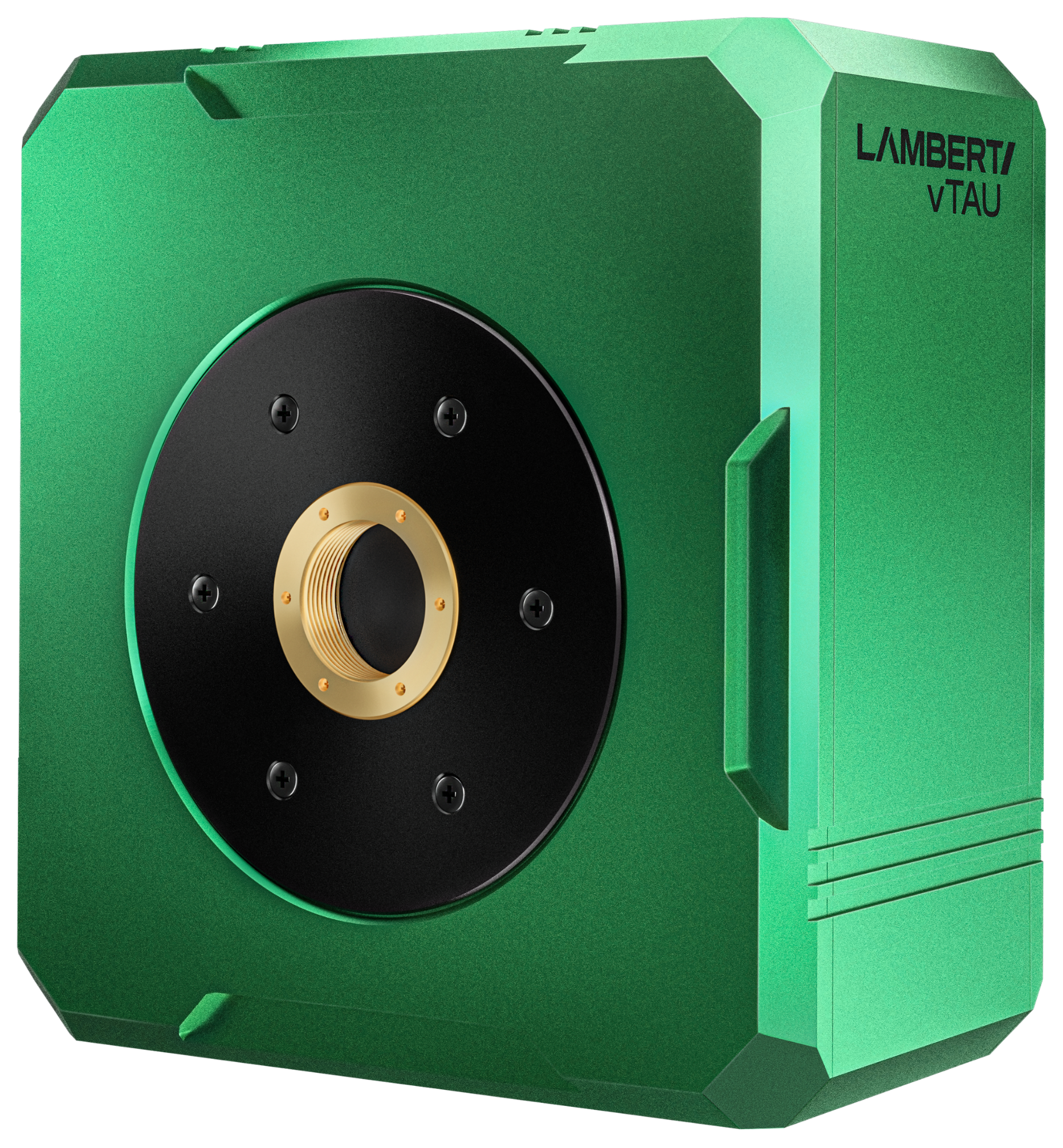
            LIFA vTAU

            SPAD powered FLIM system

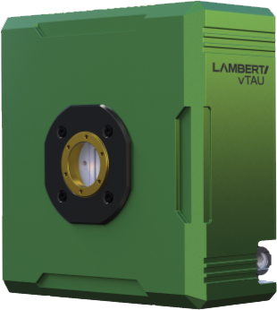
            Simplify experiments for researchers and imaging centres with the vTAU SPAD camera; combining excellent light sensitivity with easy image acquisition and data analysis.

            Minimise measurement duration, automate image acquisition, and simplify data analysis… factors of great importance for cell biology, cancer research and high-throughput screening.

            vTAU easily integrates into any FLIM system, providing a plug-n-play experience that allows for seamless switching between setups.

            Key features

            Unique image sensor

            Ultra-high sensitive SPAD detector for up to 74 FLIM images per second

            Multiple image modes

            Including regular Frequency-Domain FLIM acquisition and Time-Lapse recordings

            Characteristics

            Pixel resolution: 512 x 512 px

            Pixel size: 16 μm

            Lifetime range: 0.2 to 300ns

            Frame rate: up to 370 fps

            Sensor type: SPAD

            Downloads

            FLIM for Beginners Application Note

            Unlock a new dimension in fluorescence imaging with FLIM for Beginners. Unlike intensity-based methods, FLIM measures fluorescence decay—revealing insights into biochemical and biophysical environments. This guide covers the fundamentals, the two main approaches (Time and Frequency Domain), and Phasor Analysis, plus key applications like metabolic imaging, protein interactions, and fluorophore unmixing.

            Download it to strengthen your FLIM knowledge and enhance research in cancer, neuroscience, and drug discovery.

            System Components

            The LIFA vTAU system includes:

            Dedicated LIFA Software

            Light source(s) according to your application

            Computer with USB and HDMI connection

            On-site system installation (optional)

            1 day of hands-on training (optional)

            Phone, email and remote desktop support

            LIFA Software

            Seamless integration of hardware for full system control

            Guides user through FLIM experiments from start to finish.

            Supports third-party hardware for a flexible and expandable system.

            Time-lapse capabilities for extended experiments.

            Data export for seamless analysis and sharing.

            Triggered recording for precise data capture.

            SDK and API available for custom integrations.

            Light Sources

            Multi-LED and/or Multi-LASER as required for your set up

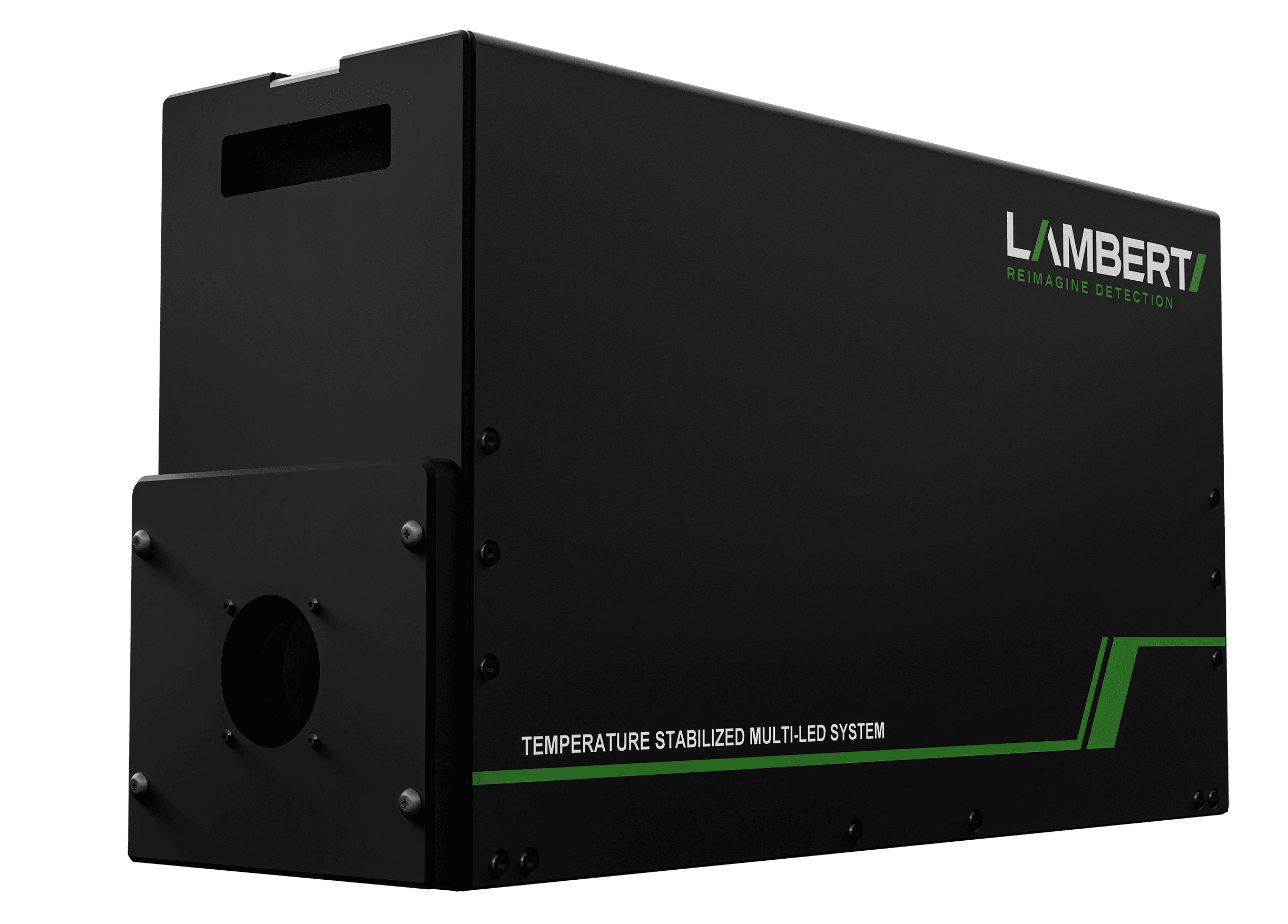
            Excitation light sources for frequency-domain fluorescence lifetime imaging, supplied according to your required wavelengths.  

            For widefield illumination, we provide multi-LED solutions.

            For laser illumination, we offer multi-LASER options with single-mode, multi-mode, and liquid light guide configurations.

            Multi-LED systems can also be provided with a liquid light guide.

            OEM Solution

            We offer an OEM platform based on the proven Lambert Instruments LIFA system

            Designed for system integrators who need a camera-based FLIM solution with short time to market.

            Our solution is compact, high-speed, and easy to integrate, giving you the flexibility to embed FLIM capabilities into your microscopy systems without compromise.

            High temporal resolution delivers fast and accurate lifetime imaging.

            Configurations

            Widefield

            On widefield microscopes, the vTAU camera in combination with the Multi-LED offers a capable yet compact FLIM solution. The vTAU is compatible with the camera port of widefield microscopes and the Multi-LED is compatible with the standard epifluorescence port of widefield microscopes.

            Spinning-disk confocal

            Being a camera-based system, the Lambert Instruments LIFA vTAU system for frequency-domain FLIM is compatible with multi-beam confocal microscopy techniques, most notably the Yokogawa CSU spinning disk series (based on the Nipkow disk scanner), Andor Dragonfly, Crest V3/V2/Cicero, and the VTInfinity series by Visitech International.

            TIRF

            Total Internal Reflection Fluorescence (TIRF) microscopy facilitates extremely high-contrast visualization and thereby high sensitivity of fluorescence near the cover glass. Typically, the optical section adjacent to the cover glass is about 100 nm. The unique combination of TIRF and frequency-domain FLIM makes it possible to measure lifetimes of, for instance, membrane receptors in order to identify their signalisation pathway. 

            Request More Information

              Lambert Instruments BV

              Leonard Springerlaan 19
              9727 KB Groningen
              The Netherlands

              User Publications

              Researchers around the world use the LIFA in their studies. Opposite is an overview of the most recent publications describing research that was done using a LIFA.

              For a complete overview of applications, please visit our applications pages and our selected LIFA publications on the Lambert main site for more information.

              Featured LIFA users

              Frequently Asked Questions

              Fluorescence Lifetime Imaging Microscopy (FLIM) is a technique that measures the decay time of fluorescence molecules, so-called fluorophores. FLIM provides detailed information about molecular environments, energy transfer between fluorophores and protein interactions. It is widely used in biological and biomedical research to study dynamic processes and cellular microenvironments.

              Frequency-domain Fluorescence Lifetime Imaging Microscopy (FLIM) is a technique used to measure the fluorescence lifetimes of fluorophores by modulating the excitation light at different frequencies. In this method, the phase shift and modulation depth of the emitted light relative to the excitation source are measured, which provides information about the fluorescence lifetime. This technique is valuable for studying molecular interactions, environment-sensitive probes, and cellular dynamics. FLIM is commonly used in biological and biomedical imaging, to sense the cellular environment, get a better understanding of cell biology or to differentiate between fluorophores based on their lifetimes if they have similar emission spectra.

              Time-domain Fluorescence Lifetime Imaging Microscopy (FLIM) is a technique that measures the fluorescence lifetime of molecules by exciting fluorophores with a very short pulse of light and then detecting the time it takes for them to emit photons. The emitted light’s intensity decays over time, and this decay curve is analyzed to determine the fluorescence lifetime.

              The primary difference between frequency-domain and time-domain FLIM lies in how the fluorescence lifetime is measured:

              • Frequency-domain FLIM: Modulates the excitation light at specific frequencies and measures the phase shift and modulation of the emitted light relative to the excitation. It determines lifetime from these shifts.
              • Time-domain FLIM: Uses short light pulses to excite fluorophores and measures the time delay between the excitation pulse and the emitted photons. The fluorescence decay curve is analyzed to determine lifetime.

              Both techniques provide insights into molecular environments but differ in how they capture fluorescence data.

              Widefield microscopy is an imaging technique used in biological and clinical research to capture images of entire specimens or large sections of tissue. It involves illuminating the entire field of view at once with light, typically from a broad light source, such as a lamp or LED. All parts of the sample are illuminated simultaneously, allowing for the detection of light emitted or reflected from the entire specimen. This method is commonly used for fluorescence microscopy, enabling researchers to observe labeled structures in live or fixed cells.

              SPAD (Single-Photon Avalanche Diode) detector technology is used to detect individual photons with extremely high sensitivity and precision. SPADs operate in “Geiger mode,” where a single photon triggers an avalanche of charge carriers, resulting in a detectable electrical pulse. This allows for the precise detection of very low light levels, making SPAD detectors ideal for applications like fluorescence lifetime imaging microscopy (FLIM), quantum cryptography, time-of-flight measurements, and biomedical imaging. SPADs offer fast response times and high photon detection efficiency, enabling high-resolution and accurate photon counting.

              Spinning-disk confocal microscopy is an advanced imaging technique used for capturing high-resolution images of biological samples in real-time. It employs a rotating disk with multiple pinholes to focus light onto specific areas of the sample while rejecting out-of-focus light. This enables faster image acquisition compared to traditional laser-scanning confocal microscopy. Spinning-disk confocal is ideal for live-cell imaging due to its speed, lower phototoxicity, and reduced photobleaching, making it suitable for dynamic processes like cell movement or protein interactions.

              Light sheet microscopy, also known as Selective Plane Illumination Microscopy (SPIM), is a technique used to image large, three-dimensional biological samples with minimal phototoxicity and photobleaching. In this method, a thin sheet of light illuminates a single plane of the sample, while an orthogonal detection system captures high-resolution images. By moving the light sheet through the sample, researchers can reconstruct detailed 3D images of tissues, embryos, or entire organisms. This method is especially useful for long-term live imaging of biological processes in real-time.

              Total Internal Reflection Fluorescence (TIRF) microscopy is a technique used to observe events occurring near the surface of a cell, typically at or near the plasma membrane. TIRF systems use an evanescent wave generated by light undergoing total internal reflection at the interface between two media (e.g., glass and water) to selectively illuminate and excite fluorophores within a very thin region, typically less than 200 nm, adjacent to the surface. This minimizes background noise and allows high-contrast imaging of cellular processes like protein interactions at the cell membrane.

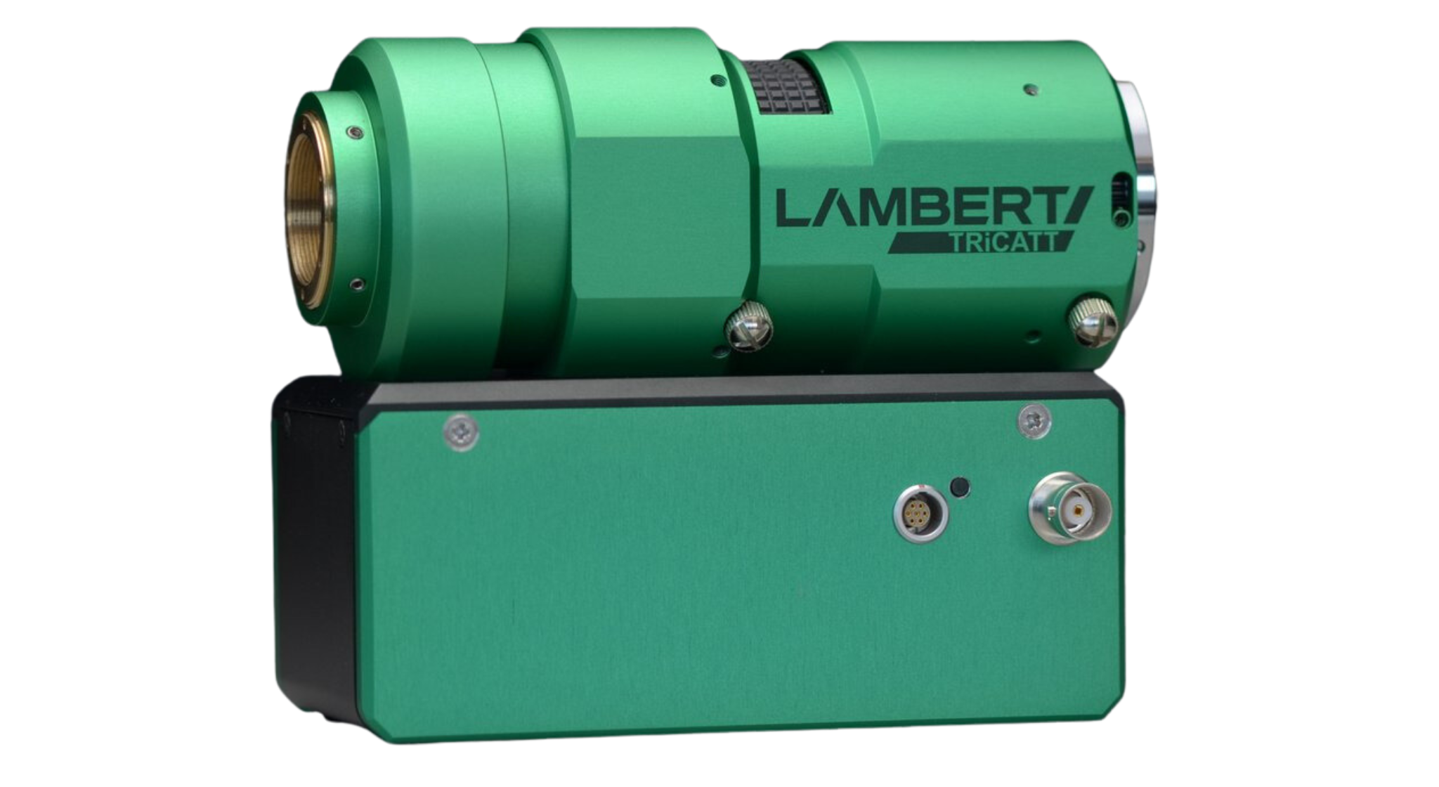
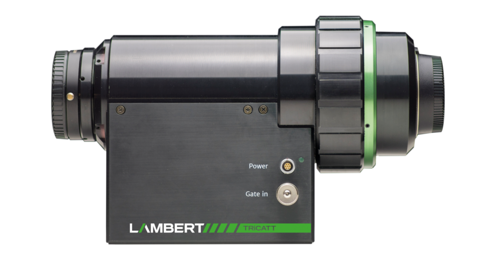
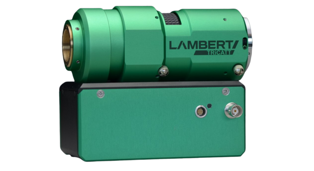
              TRiCATT

              Compact lens-coupled image intensifier

              TRiCATT

              HIGH RESOLUTION

              Gen II and Gen III image intensifiers offering the world’s highest resolution and sensitivity in the UV, visible or near infrared

              IN-SITU FOCUS ADJUSTMENT

              outer-body focus ring adjusts the internal relay optics without altering total length

              COMPACT DESIGN

              easily fits within your imaging or spectroscopy setup, with a super-compact 18mm version also available 

              Description

              The TRiCATT is a compact lens-coupled image intensifier for scientific and industrial applications that require:

              – Low-light level imaging

              – Ultra-short exposures through fast gating

              Any camera with C-mount and a 1/2″, 2/3″, or 1″ image sensor is compatible with the TRiCATT. You can find the right TRiCATT for your camera with our interactive calculator.

              Applications

              Time-resolved imaging and spectroscopy

              Particle Image Velocimetry (PIV)

              Laser Induced Fluorescence (LIF)

              Diffuse Optical Tomography (DOT)

              Time-gated luminescence

              Forster Resonance Energy Transfer (FRET)

              Oxygen imaging

              Viscosity imaging

              Single-molecule imaging

              Bio- and chemiluminescence imaging

              Solar PV and LED characterization

              Combustion research

              Time-gated Raman

              Plasma physics

              X-ray Imaging

              Single photon imaging

              Features

              Small gate widths
              Gate width down to less than 3 ns (FWHM) with minimal jitter.

              High gate repetition widths
              Up to 300 kHz / 2.5 MHz burst. 1Mhz Optional

              Overexposure Protection
              User-definable current limitation and optional shutter.

              Easy Coupling
              Efficient lens coupling to any CCD and CMOS camera (up to 500 fps) with C-mount input and output.

              Relay Lens
              The high quality relay lens transfers the intensified image to the image sensor of the attached camera very efficiently and without losses in resolution. If required we can provide the 0.5x relay lens with a back focal distance of 13 mm.

              Camera
              Along with the Image Intensifier TRiCATT Lambert Instruments can deliver different types of CCD and CMOS cameras. If you already have a camera, you can use our interactive calculator to determine which intensifier size and relay optics are best suited for your setup.

              Match my camera

              The relay optics of HiCATT and TRiCATT project the output of the image intensifier onto the sensor of your camera. This calculator helps you determine the optimal configuration of image intensifer diameter and relay optics for your camera and application.

              Read More

              Fluorescence Imaging

              Image Intensifiers 

              An image intensifier boosts the intensity of the incoming light. By converting photons to electrons and back to photons, the light intensity can be increased significantly. Scroll down for more information about how this works.

              Another feature of an image intensifier is that it can act as an ultra-fast shutter. The photocathode can be switched between on and off in a matter of nanoseconds.

              Read More

              Fluorescence Imaging

              Photocathodes 

              The photocathode is the entrance of the image intensifier. This is where the incoming photons are converted to electrons. The quantum efficiency of the photocathode material specifies how efficient this conversion is for each wavelength.

              Read More

              Phosphors

              The output of the image intensifier contains a layer of phosphorescent material. Upon impact of an electron, the phosphor screen will emit light. Depending on the type of phosphor, the intensity of the emitted light will decrease faster.

              Read More

              Specifications and Options

              HiCATT Image Intensifier

              TRiCATT 25

              Image intensifier: 25 mm image intensifier. Gated or modulated. P43 phosphor

              Minimum gate time: 3 ns, 40 ns or 50 ns

              Gate repetition rate*: 100 kHz, 300 kHz or 1 MHz

              Relay optics**: 1:1 or 1.7:1 Fixed aperture

              Input mount: F-mount and C-mount

              Output mount: F-mount and C-mount

              Mechanical shutter: Optional

              Mounting plate: Optional

              TRiCATT 18

              Image intensifier: 18 mm image intensifier. Gated or modulated. P43 phosphor

              Minimum gate time: 3 ns, 40 ns or 50 ns

              Gate repetition rate*: 100 kHz, 300 kHz or 1 MHz

              Relay optics**: 1:1 Fixed aperture

              Input mount: F-mount and C-mount

              Output mount: F-mount and C-mount

              Mechanical shutter: Optional

              Mounting plate: Optional

              TRiCATT

              TRiCATT 18C

              Image intensifier: 18 mm image intensifier. Gated or modulated. P43 phosphor

              Minimum gate time: 3 ns, 40 ns or 50 ns

              Gate repetition rate*: 100 kHz, 300 kHz or 1 MHz

              Relay optics**: 1:1 Variable aperture

              Input mount: C-mount

              Output mount: C-mount

              Mechanical shutter: Custom design upon request

              Mounting plate: Optional

              Gating Specifications

              Standard Gating

              Width range: 40 ns – 10 s

              Resulting min. pulse width (increments): 40 ns (20ns)

              Pulse repetition rate: < 10 MHz

              Delay jitter (width): ± 10 ns (± 250 ps)

              Insertion delay: 100 ns

              Fast Gating

              Width range: < 3 ns – 10 s

              Resulting min. pulse width (increments): < 3 ns (10 ps)

              Pulse repetition rate: < 16 MHz

              Delay jitter (width): < 35 ps (< 35 ps)

              Insertion delay: 20 ns

              Trigger input: Programmable trigger level, divider and bursts (m out of n triggers)


              Control Units

              The control unit contains a micro-controller, a high voltage power supply and a RF (Radio Frequency) amplifier. The control unit has a low voltage input to receive the external modulation signal. It amplifies this signal and biases it with a variable DC photocathode voltage. The control unit offers control of the MCP voltage for setting the image intensifier gain. The control unit also monitors the light output, and switches off the image intensifier when its light output becomes too high. The control unit supports modulation frequencies up to 120 MHz.


               Manual Gain ControllerGain ControlGate ControlGate Generator
              Gain control   
              Anode current limiter  
              Shutter control  
              Gain control 
              Gate control  
              Internal trigger generator  
              Programmable gate   

              Options

              Optional: Signal Generator
              Instead of using an external modulation signal generator, we offer a built-in modulation signal generator as part of the control unit/power supply for frequencies up to 120 MHz.

              Optional: TRiCAM
              As an alternative to the lens-coupled ICCD camera (TRiCATT + CCD), we offer an ICCD camera in which the image intensifier is fiber-optically coupled to the sensor. This is the TRiCAM. This modulated intensified CCD camera is very compact and has a significantly higher gain than the lens-coupled combination as a result of the more efficient and compact fiber coupling.

              Downloads

              For more information on the TRiCATT please download our brochure.

              Request More Information

                Lambert Instruments BV

                Leonard Springerlaan 19
                9727 KB Groningen
                The Netherlands

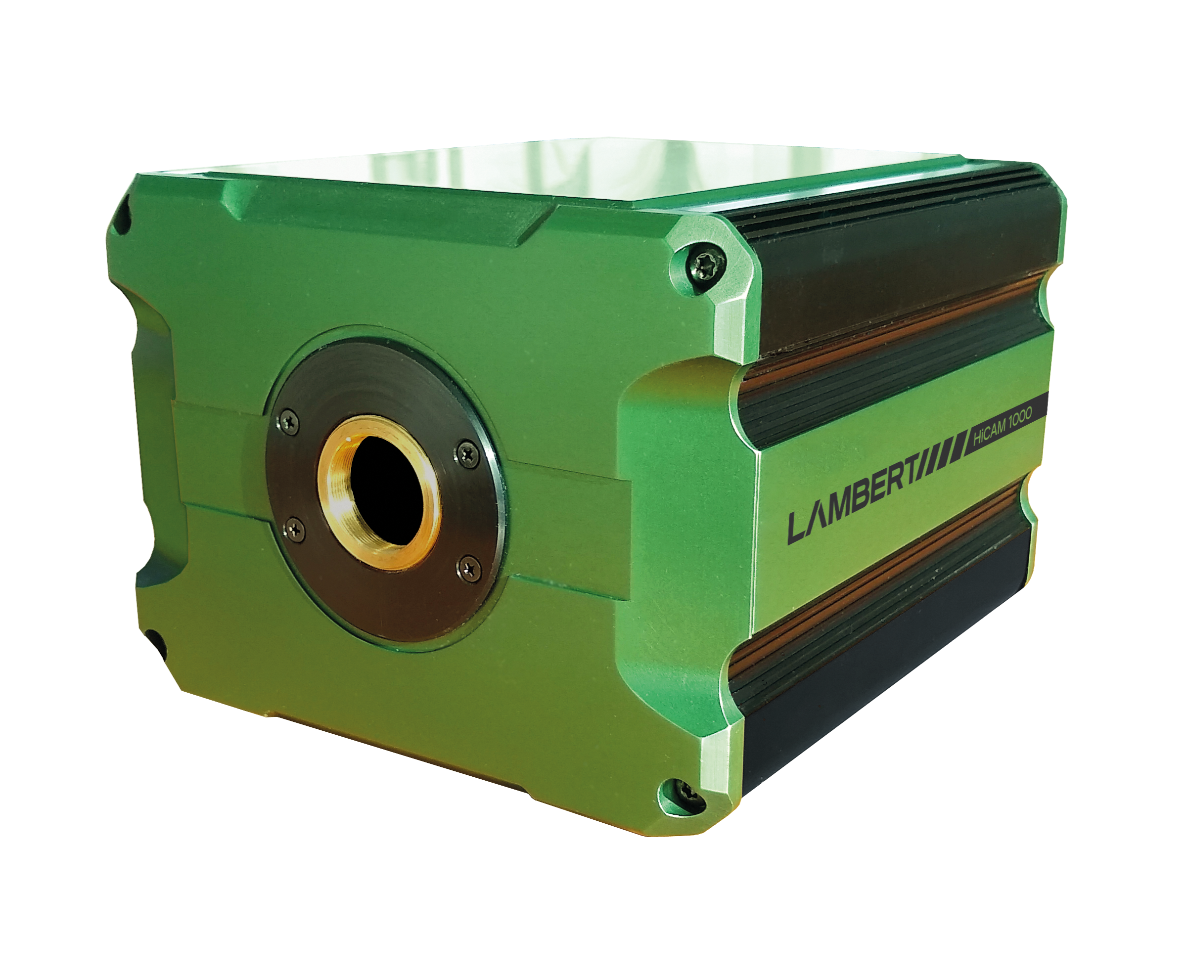
                HiCAM

                Intensified high-speed high-sensitivity imaging camera

                Fluorescence Microscopy Cameras

                HIGH PERFORMANCE

                delivering unprecedented stamina, speed and sensitivity

                HIGH SPEED

                single photon counting suitable for particle physics by making use of scintillation

                HIGH SENSITIVITY

                ranging from Deep UV to NIR to match the spectral needs of your application

                Description

                The HiCAM is a gated intensified high-speed high-sensitivity imaging camera. It has an integrated fiber-optically coupled image intensifier, which offers a unique combination of high speed and sensitivity down to single photon level. Because the HiCAM does not need high intensity light sources, it is suitable for use in low-light level conditions.

                Applications

                Super-slow motion combustion research for the automotive industry

                Time-resolved imaging in plasma physics research

                Dynamic phenomena in microscopy, e.g. Imaging the rotation of single molecules of ATPase

                Laser-Induced Fluorescence (LIF)

                Flow visualization and velocity measurements using Particle Image Velocimetry (PIV)

                Time-resolved imaging of fluids for microfluidic research

                Blood flow analysis

                Time-resolved fluorescence

                UV imaging for hydrogen combustion

                Features

                Ultra-short Gating
                The camera’s effective exposure time can be reduced to < 3 ns (FWHM) for time resolved imaging, or capturing very fast events.

                Fast Streaming
                To transfer all the high-resolution image data, the HiCAM streams live over a CoaXPress (CXP) interface. The camera has four CXP connectors, each of which has a channel speed of 6.25 Gbit/s.

                Longterm Streaming
                The recordings can be directly streamed to the hard drive. For optimised recorders the length is limited only by drive capacity.

                High-sensitivity Intensifiers
                You can choose from a wide variety of high-sensitivity image intensifiers to match the spectral needs of your application.

                Cycled Bursts
                Adjust for changing light levels by cycling through three different gate widths and optionally combine this with the burst mode to optimize the dynamic range.

                Fiber-optically Coupled
                Our experienced engineers couple the sensor to the image intensifier with a fiber-optic window.

                Fluorescence Imaging

                Image Intensifiers 

                An image intensifier boosts the intensity of the incoming light. By converting photons to electrons and back to photons, the light intensity can be increased significantly. Scroll down for more information about how this works.

                Another feature of an image intensifier is that it can act as an ultra-fast shutter. The photocathode can be switched between on and off in a matter of nanoseconds.

                Read More

                Fluorescence Imaging

                Photocathodes 

                The photocathode is the entrance of the image intensifier. This is where the incoming photons are converted to electrons. The quantum efficiency of the photocathode material specifies how efficient this conversion is for each wavelength.

                Read More

                Videos

                Super-slow motion video of a gas flame recorded with the Lambert Instruments HiCAM at 1000 fps (frame rate) and gating of 15 us (effective exposure time). Resolution: 1280 x 512 pixels.

                High-speed recording of a cork burning in plasma. Recorded with a HiCAM at 5000 fps.

                Recording of a beating heart of a zebrafish. Recorded at 2000 fps with the HiCAM on a fluorescence microscope. The blood cells were stained with a DS-red fluoropho.

                A touch-me-not plant bursting open. Captured with a HiCAM with 18 us gating.

                Specifications

                HICAM, 1000 

                Framerate (Full Resolution): 1000 fps

                Sensor Resolution: 1280 x 1024 pixels

                Streaming: HiCAM 1000: CoaXPress

                Bit Depth: 8 and 12 bit

                HICAM, 2000 

                Framerate (Full Resolution): 2000 fps

                Sensor Resolution: 1920 x 1080 pixels

                Streaming: HiCAM 2000: CoaXPress

                Bit Depth: 8

                The dual-stage image intensifier, specially designed for high-speed cameras, can be equipped with a variety of photocathodes; ranging from ultra violet to infra red. Adapting the photocathode will give maximum output brightness to enhance Signal-to-Noise Ratio (SNR). Moreover, the image intensifier is fiber-optically coupled to a CMOS sensor. This further increases SNR, as compared to a lens-coupled system.

                Downloads

                For more information on the HiCAM, please download the brochure.

                Request more information

                  Lambert Instruments BV

                  Leonard Springerlaan 19
                  9727 KB Groningen
                  The Netherlands

                  User Publications

                  Researchers around the world use the HiCAM for high-speed high-sensitivity imaging in their studies. Opposite is an overview of the most recent publications describing research that was done using a HiCAM.

                  For a complete overview of applications, please visit our applications pages.

                  STAMINA

                  Complete high-speed recording solution

                  high-speed recording with multiple cameras

                  ZOOM BACK INTO TIME

                  to optimise efficiency and reliability of your production process

                  BACK TO BACK RECORDINGS

                  without intermittent downloads

                  MULTI-CAMERA SYSTEM

                  view and record images with multiple cameras at the same time.

                  Description

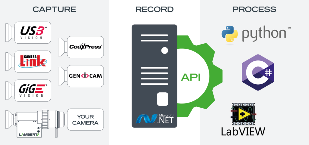
                  Stamina combines advanced hardware with easy to use software to offer a complete multi-camera recording solution for your imaging challenges. With the complete imaging system it becomes possible to record, view and combine multiple cameras in one setup. Our software is compatible with a wide range of cameras, providing the option to integrate your own cameras into the imaging setup easily.

                  With Stamina, all recordings can be synchronised and all image data is directly streamed to the solid storage, enabling longterm streaming. With the external Application Programming Interface (API), Stamina can be further customised and integrated in your existing setup. Through the API, you have access to device settings, recording parameters and image data.

                  Stamina is a completely customizable multi-camera recording and imaging solution. Our experienced sales engineers will advise you on the right system for your application.

                  Applications

                  Troubleshooting high speed events (triggered recording)

                  Reliability engineering

                  Long-term multi camera recording

                  High speed / High resolution recording with multiple cameras

                  Features

                  Add your Own Camera
                  Stamina is compatible with a wide range of cameras, providing the option to integrate your own (high-speed) cameras into the imaging setup easily.

                  Complete Solution
                  Stamina combines advanced hardware with easy to use software to offer a complete solution for your imaging challenges.

                  Long term streaming
                  By directly streaming to the hard drive, Stamina makes long term streaming easy. The maximal recording length only depends on the available storage space.

                  Control all hardware into one place
                  Change and control the settings of all compatible hardware in one place with Capture.

                  API Available
                  Integrate Stamina into your existing setup with the Application Programming Interface (API). The following platforms are compatible with the API: MATLAB, LabVIEW, Python 3 (64 bit).

                  Video

                  System set up

                  Multi-Camera Recording

                  Downloads

                  For more information on the STAMINA please download our brochure.

                  Request more information

                    Lambert Instruments BV

                    Leonard Springerlaan 19
                    9727 KB Groningen
                    The Netherlands

                    News Publications

                    Read about STAMINA’s launch at Photonics West 2022.

                    HiCAM Fluo

                    Cooled High-speed Camera for Fluorescence Imaging

                    High-Speed Camera

                    HIGH SPEED CAMERA

                    frame rate over 2000 fps

                    HIGH SENSITIVITY

                    down to single photon level

                    LONG TERM STREAMING

                    record directly to hard drive

                    Description

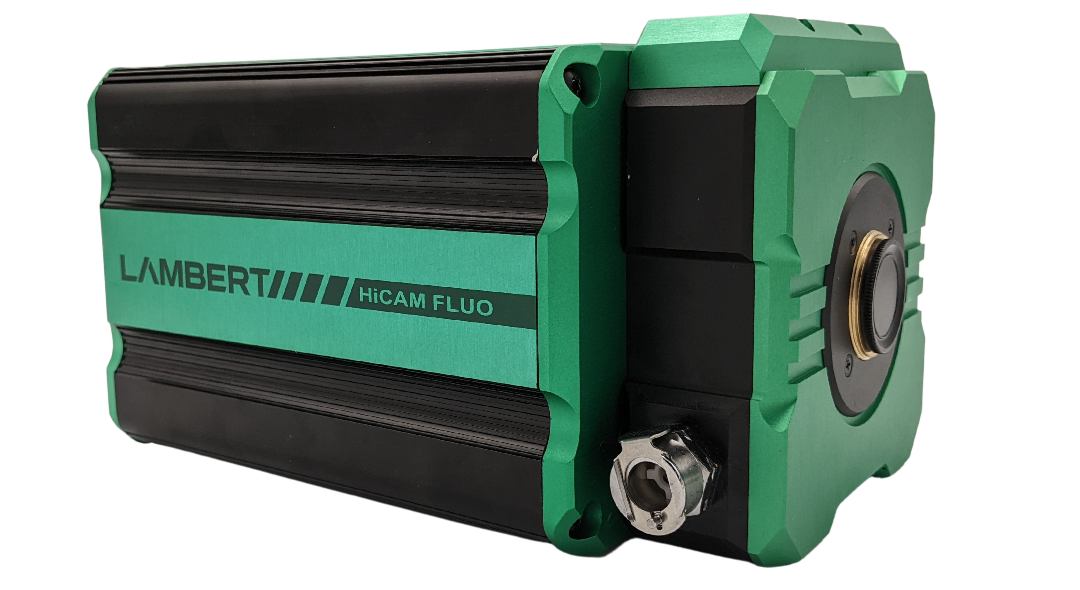
                    The HiCAM Fluo is a high-speed camera for fluorescence imaging applications. It records high resolution images at a frame rate over 2000 fps in the most challenging low-light conditions by using a cooled image intensifier. Packed into a compact aluminum enclosure, it is easy to attach the HiCAM Fluo to any fluorescence microscope. 

                    Fluorescence Imaging Applications

                    High-speed fluorescence imaging, bioluminescence and chemiluminescence detection for in-vivo imaging

                    Time-resolved imaging of fluids for microfluidic research

                    Particle Image Velocimetry (PIV)

                    Time-resolved imaging and spectroscopy using ultra-short exposures (<3ns)

                    Laser-Induced Fluorescence (LIF)

                    Super-slow motion combustion research

                    Plasma physics research

                    ·       Single-photon imaging microscopy and quantum physics 

                    Plasma physics research for fusion energy

                    Features

                    Fiber-optically coupled intensifier
                    This offers a unique combination of high speed imaging and increased light sensitivity down to single photon level. 

                    CoaXPress
                    To transfer all high-resolution image data, the HiCAM Fluo streams live over a CoaXPress (CXP) interface. The camera features four CXP connectors, each supporting channel speeds of up to 6.25 Gbit/s (CXP-6). For higher bandwidth requirements, CXP-12 compatibility ensures even faster data transfer, optimizing performance for demanding imaging applications.

                    Ultra-Short Gating
                    With its gated image intensifier, the camera’s effective exposure time can be reduced. The minimum gate width of 10 ns (FWHM) increases the range of light levels at which the camera can be used. It also eliminates motion blur and enables time-resolved filtering.

                    Cooled Image Intensifier 
                    The fanless design of the camera minimises vibrations to ensure sharp images. Very low noise levels are achieved by Peltier cooling the image intensifier. Noise levels are reduced by a factor of up to 100 times as compared to uncooled intensified cameras.
                      

                    Image Intensifiers 

                    An image intensifier boosts the intensity of the incoming light. By converting photons to electrons and back to photons, the light intensity can be increased significantly. Scroll down for more information about how this works.

                    Another feature of an image intensifier is that it can act as an ultra-fast shutter. The photocathode can be switched between on and off in a matter of nanoseconds.

                    Read More

                    Fluorescence Imaging

                    Photocathodes 

                    The photocathode is the entrance of the image intensifier. This is where the incoming photons are converted to electrons. The quantum efficiency of the photocathode material specifies how efficient this conversion is for each wavelength.

                    Read More

                    Fluorescence Imaging Videos

                    Recording of a beating heart of a zebrafish. Recorded at 2000 fps with the Lambert Instruments HiCAM on a fluorescence microscope. The blood cells were stained with a DS-red fluorophore.

                    monArch fluorescent voltage indicator observed with the Lambert Instruments HiCAM Fluo (1000 fps, 1200 x 1000 pixels).

                    SCAPE 2.0 dual-color, high-speed imaging of blood flow in a beating embryonic zebrafish heart acquired at 100 volumes per second over a 270 x 260 x 128 xyz micron field of view. The heart wall is shown in green (EGFP) and the red blood cells are shown in red (DsRed). Collaboration between Hillman Lab and Kimara Targoff.

                    Specifications

                    HICAM FLUO, 1000 FPS 

                    Maximum resolution: 1280 x 1024 pixels

                    Frame rate:

                    1000 fps at full resolution

                    1500 fps at 1200 x 720 pixels

                    4000 fps at 640 x 480 pixels

                    7500 fps at 256 x 256 pixels

                    Pixel size: 6.6 um 

                    Pixel depth: 8 or 12 bit

                    Minimum exposure time: 40 ns

                    Gating repetition rate: 100 kHz 

                    Image intensifier: GaAsP (Others optional)

                    Computer interface: Streaming CoaXPress

                    Photon gain (max.): 36000 lm/m^2/lx


                    HICAM FLUO, 2000 FPS 

                    Maximum resolution: 1920 x 1080 pixels

                    Frame rate: 

                    2000 fps at full resolution

                    2500 fps at 1280 x 960 pixels

                    5100 fps at 640 x 480 pixels

                    6400 fps at 512 x 384 pixels

                    Pixel size: 10 um

                    Pixel depth: 8 bit 

                    Minimum exposure time: 40 ns

                    Gating repetition rate: 100 kHz

                    Image intensifier: GaAsP (Others optional)

                    Computer interface: Streaming CoaXPress 2.0

                    Photon gain (max.): 36000 lm/m^2/lx

                    Downloads

                    For more information please download the brochure or datasheet.

                    Request more information

                      Lambert Instruments BV

                      Leonard Springerlaan 19
                      9727 KB Groningen
                      The Netherlands

                      User Publications

                      Researchers around the world use the HiCAM Fluo in their studies. Opposite is an overview of the most recent publications describing research that was done using a HiCAM Fluo.

                      For a complete overview of applications, please visit our applications pages.

                      Contact Us

                      5th floor,
                      Leonard Springerlaan 19
                      9727KB Groningen
                      The Netherlands

                      Phone:
                      +31 (0) 50 501 8461
                      Email:
                      sales@lambertinstruments.com